At IMB, we explore the building blocks of life itself in remarkable and unprecedented detail for disease discovery, invention and application.
Using cutting-edge research technologies and facilities, our researchers generate powerful insights and essential information to understand, prevent and treat disease; and create more sustainable cities, fuels and foods.
This year's IMB Science as Art Awards, which are an initiative of the Students of IMB Association (SIMBA), provide an opportunity for us to share the beauty and wonder of the building blocks of life through images and artworks that excite and inspire us.
Please take a few minutes to explore the stunning images and insightful stories below created by our scientists for this year's awards and view the award winners.
The haze is purple - Winner (Imaging)
This is an image of an immune cell called a macrophage in the process of engulfing foreign particles. The image was composed from a raw data set taken from the 3View electron microscope at UQ. Our lab focuses on understanding the roles of key players in the inflammatory response. Our approaches are designed to understand the key host cell targets for the future development of new anti-microbial drugs and vaccines.
Image: Darren Brown | Related content: Stow Group | Centre for Inflammation and Disease Research
Amazing zebrafish - Runner-up (Imaging)
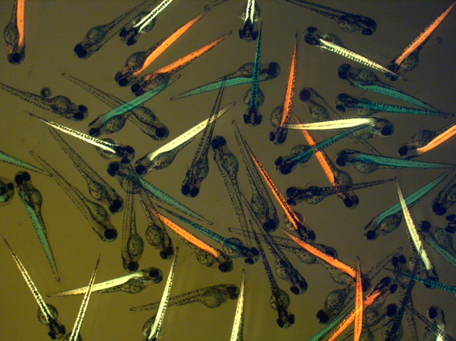
This image shows zebrafish embryos from outcrossing Cavin-GFP fish with APEX fish: the green embryos were expressing green fluorescent protein (GFP)- tagged Cavin protein, the red embryos were APEX transgenic fish expressing red fluorescent protein, and the yellow embryos were expressing both CavinGFP and APEX. The non-fluorescent embryos did not expressing either transgenes.
We were attempting to develop a novel in vivo proteomic-mapping platform in zebrafish. This platform uses a GFP-directed APEX biotinylation system, and this system can be applied for screening interactors of the Cavin, a protein family that regulates caveolae formation and involved in muscle development and muscular dystrophies. These transgenic fish were used to reveal Cavin-associated signalling pathway and the molecular mechanism underlying human muscular dystrophies.
Image: Zherui Xiong | Related content: Parton Group | Microscopy facility
Grandiflora - Winner (Multimedia)
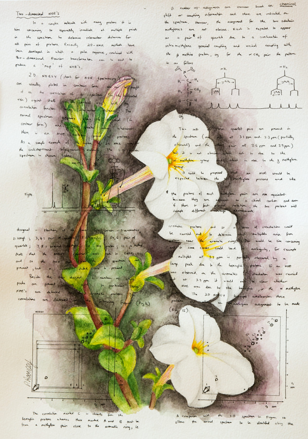
Researchers at IMB’s Clive and Vera Ramaciotti Facility for Producing Pharmaceuticals in Plants are establishing systems to transform plants into ‘biofactories’. In this illustration, the background of handwritten lecture course notes refers to the knowledge base and education that benefit from basic scientific research. From this strong foundation grows applications and remedies for the future. Plants such as canola, tobacco and petunia (as shown) are being used to express compounds of interest for mass production of next-generation pharmaceuticals to treat a range of conditions such as cancer, pain and obesity.
Image: Dr Peta Harvey | Related content: Craik Group | Clive and Vera Ramaciotti Facility for Producing Pharmaceuticals in Plants
Bacteria caught in the act - Runner-up (Multimedia)
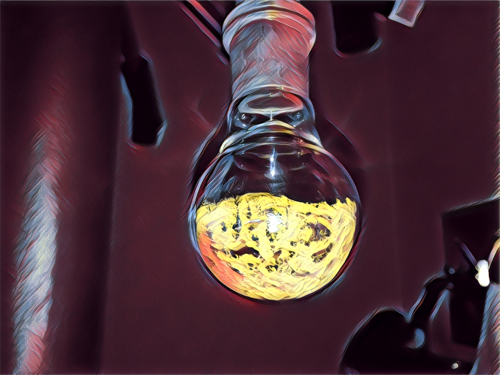
Riboflavin is an essential vitamin for humans and bacteria, but did you know that humans obtain it whole from the diet, while bacteria construct riboflavin from scratch? After a decade-long search, we found that our immune system can sense the presence of bacteria by detecting their half finished riboflavin molecules, like unfinished cars in a factory production line. Obtaining these molecules is extremely important for immunological research in order to understand how our immune system detects and fights bacteria. Unfortunately, these molecules are unstable and degrade within minutes. So, in order to 'trap' these molecules for research use, we developed special chemical techniques and methods. This stylised photo shows our successful trapping of these highly unstable molecules as a bright yellow powder. This chemical has already enabled researchers worldwide to gain key immunological insights, and may form the basis of future vaccines or autoimmune disease targeting drugs.
Image: Dr Jeffrey Mak | Related content: Fairlie Group | Nuclear Magnetic Resonance (NMR) Spectroscopy Facility | Centre for Inflammation and Disease Research
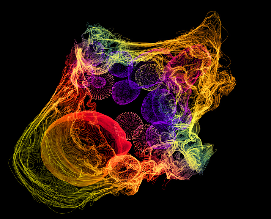
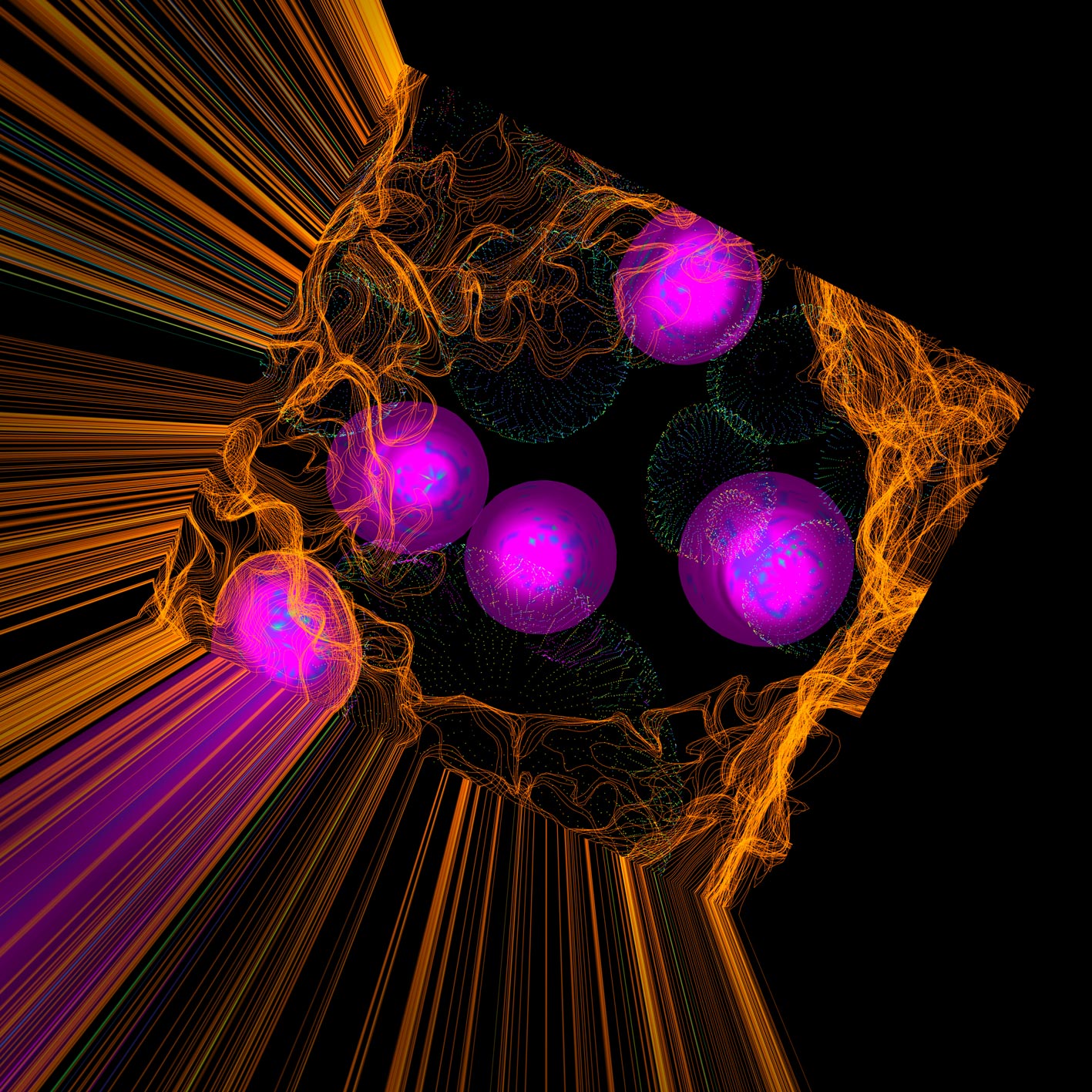
Earth's solar interface - Runner-up (Multimedia)
Earth receives 2600x more solar energy from the sun than we need to power our entire global economy. Higher plants and single-cell green microalgae have evolved over billions of years to tap into the huge energy source of the Sun, and to use it to power the process of photosynthesis which captures and fixes CO2, producing atmospheric oxygen and biomass which consists of a complex set of biomolecules that collective provide us with food, fuel and a wide range of high value chemical feedstocks.
This is a multiscale movie focused on microalgae systems. In this short movie, we zoom into these systems from the planetary scale down to the atomic detail of the photosythtetic machinery that powers these solar driven microalgae technologies. This multiscale analysis not only illustrates the intrinsic beauty of these complex systems but also enables structure-guided design of next-generation microalgae technologies.
Video: Rohan Drysdale | Related content: Hankamer Group | Centre for Solar Biotechnology
Back to the lab - Winner (Science Communication)
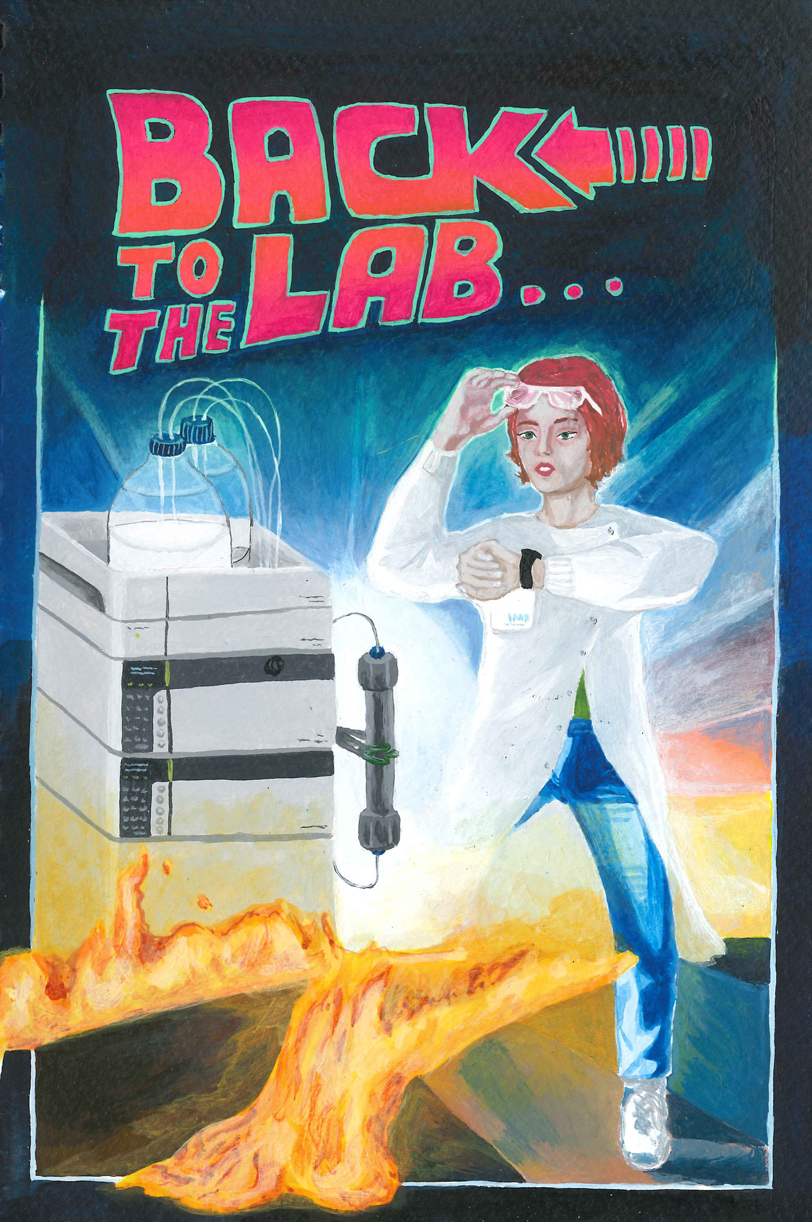
Going back in time and back to the future, e.g. to save an experiment, is something many PhD students wish they could do. Usually, they just go back to the lab. Picture is based on the 1985 movie poster "Back to the Future".
Image: Felicitas Vernen | Related content: Craik Group
Standing out - Winner (People's Choice Award)
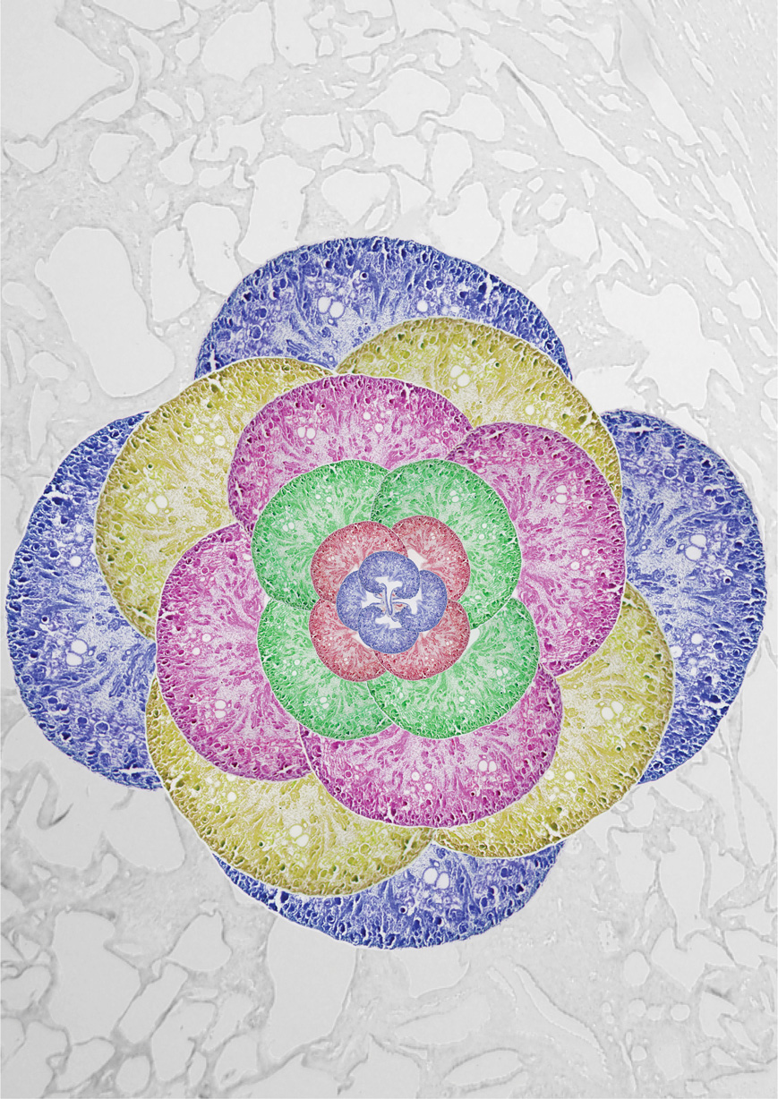
In the image, we can see pseudocolored sagittal kidney sections. To understand many biological processes, in our lab we have access to different tools to visualise what is happening inside the organs of different organisms. Being able to observe so closely the morphology of certain organs gives us a clearer insight and understanding on their function.
Image: Maria Rondon Galeano | Related content: Hogan Group | Microscopy facility
Inflamed
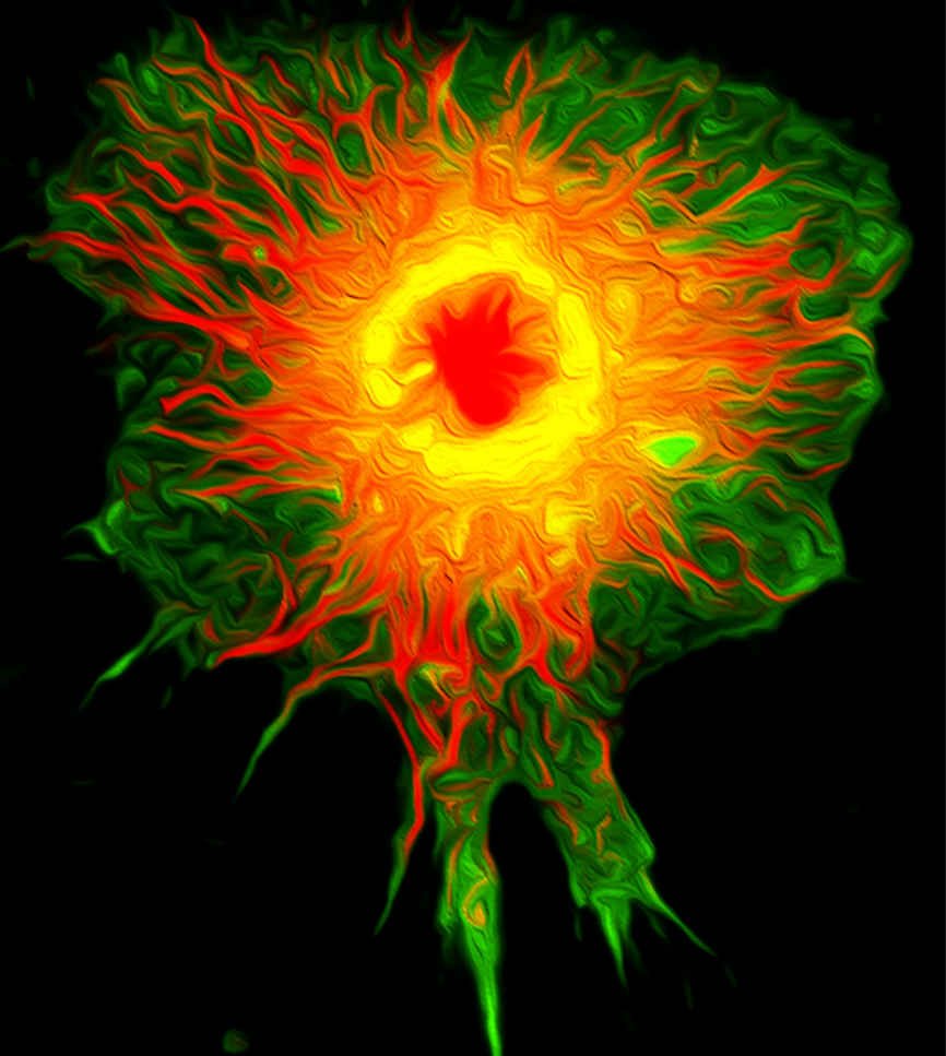
Nothing says inflammation like this fired up macrophage! Activated to produce the inflammatory cytokine TNF (shown in green) which is being made in the Golgi complex (shown in yellow) and is pouring out of the cell along microtubule highways (shown in red), this macrophage will be soon be killing bacteria and causing heat and pain just to let us know it's on the attack.
Image: Professor Jenny Stow | Related content: Stow Group | Microscopy facility | Centre for Inflammation and Disease Research
Diffraction
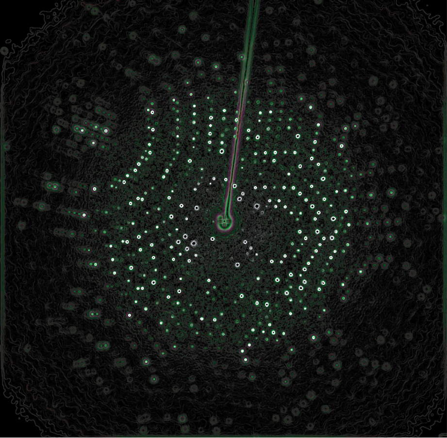
This is a diffraction image from an X-ray crystallography experiment. X-ray crystallography is a technique used by IMB researchers to determine what molecular machines look like, or their ‘structure’. After growing crystals of a molecular machine, researchers shoot them with an X-ray beam. If the molecules inside the crystal line up well with each other, this experiment will result in “diffraction”. These dotted patterns hold the information needed by researchers to determine what the molecular machine looks like. Researchers use a number of computer programs that make the process of converting diffraction into a 3-dimensional structure much easier. Knowing the structure of a molecular machine can help us understand disease and biological processes or even assist in drug discovery.
Image: Emily Furlong | Related content: Collins Group | UQROCX Crystallisation Facility
Kidney arrangement
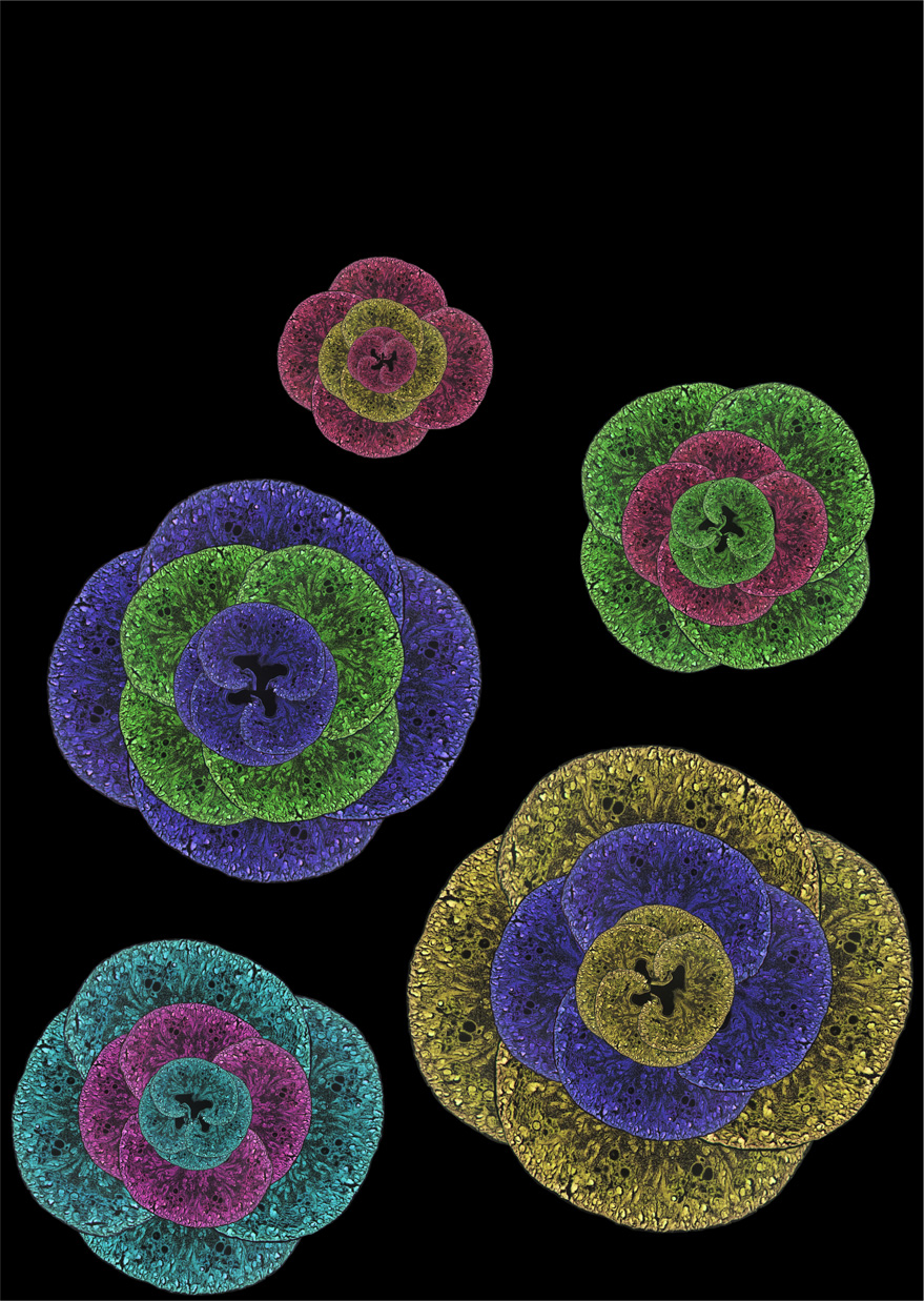
Arrangement of pseudocolored sagital kidney sections. To understand many biological processes, in our lab we have access to different tools to visualise what is happening inside the organs of different organisms. Being able to observe so closely the morphology of certain organs gives us a clearer insight and understanding on their function.
Image: Maria Rondon Galeano | Related content: Hogan Group | Microscopy facility
Conus
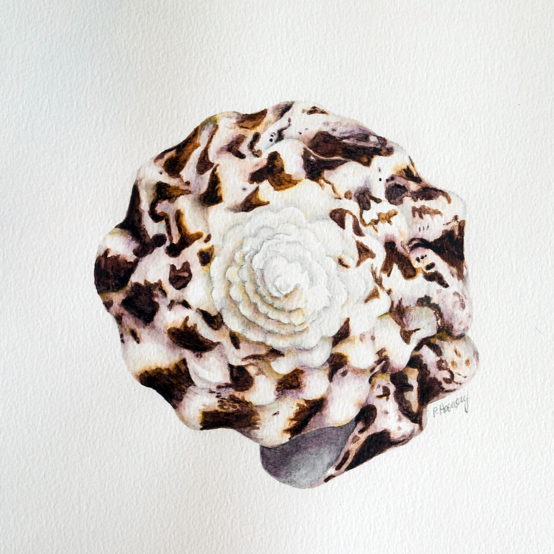
Brightly coloured and patterned, it is not merely the beauty of cone shells that attracts scientists to work with these predatory sea snails. Rather it is their venom, consisting of a cocktail of peptides known as conotoxins. These compounds are highly effective and selective for a range of ion channels and receptors. This activity gives these peptides excellent potential as lead molecules in drug design, especially as analgesics. Researchers within IMB are working with a conotoxin from Conus regius with the aim of better understanding how these peptides may help in the treatment of chronic neuropathic pain.
Image: Dr Peta Harvey | Related content: Craik Group | Centre for Pain Research
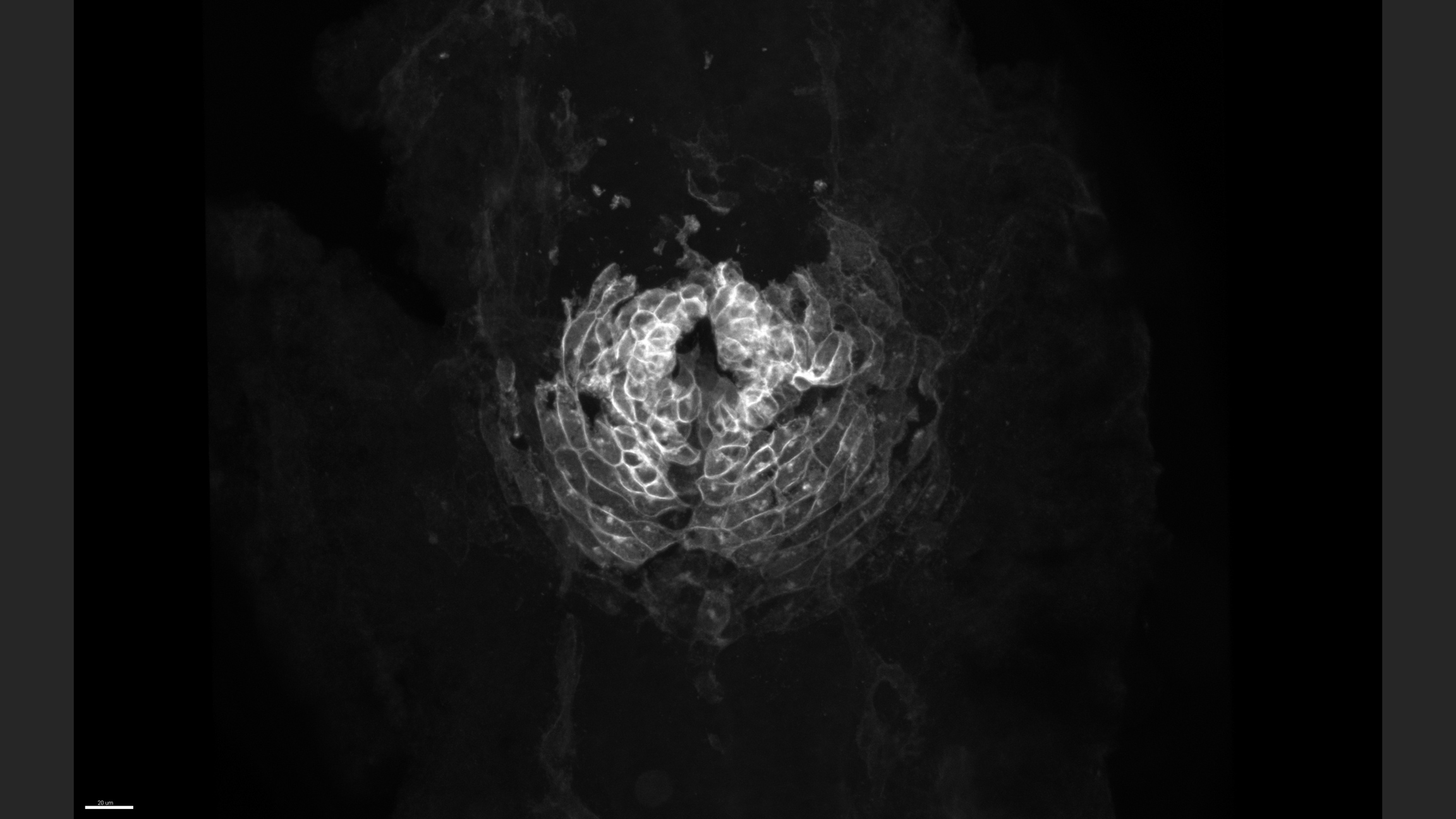
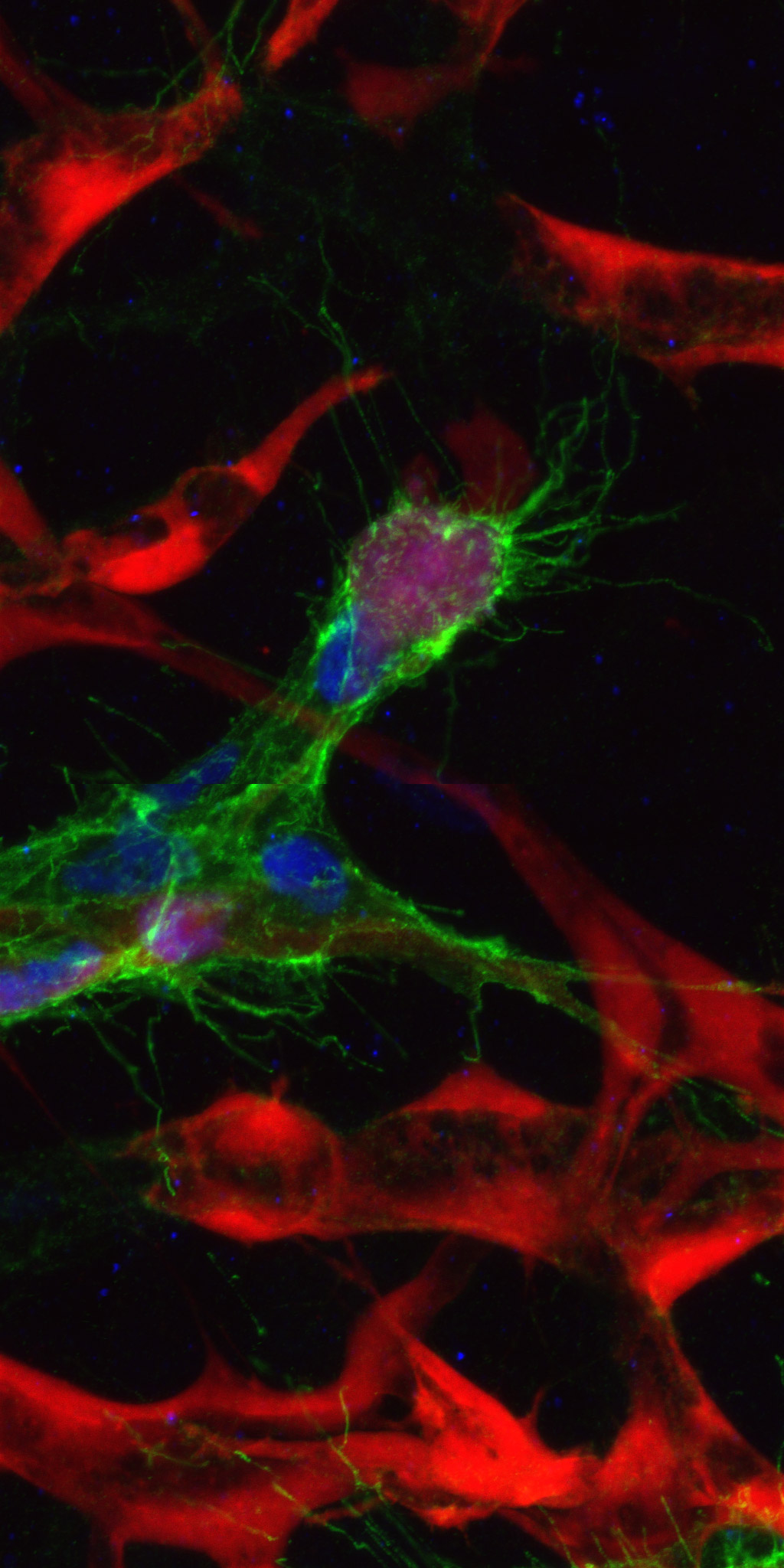
Migrating lymphatics
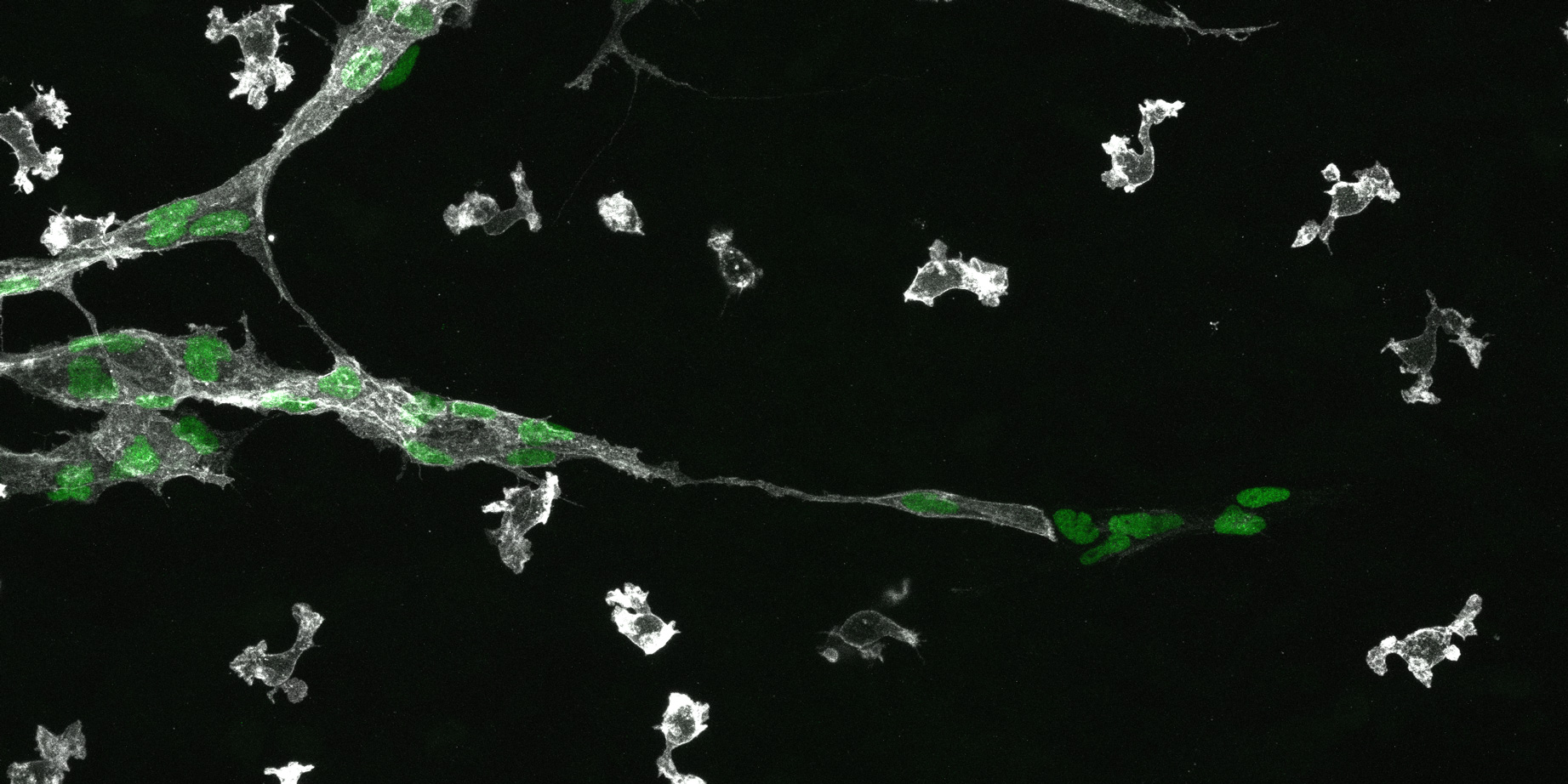
This image is showing a migrating lymphatic vessel during development. The nuclei of the cells forming in this lymphatic vessel are visible in green and the membrane of cells in grey. You can also see some macrophages (isolated cells in grey).
Image: Dr Cathy Pichol-Thievend | Related content: Francois Group | Microscopy facility | Centre for Cardiac and Vascular Biology
Beauty in the beast
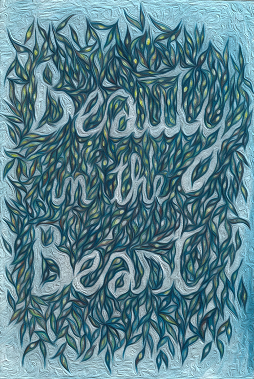
My research focuses on peptides with anticancer properties and I am working with various cancer cell lines. While cancer is still one of the leading causes of death worldwide, cancer cells grown in cell culture flasks (on plastic) can be quite beautiful to look at.
Image: Felicitas Vernen | Related content: Craik Group
Christmas lights
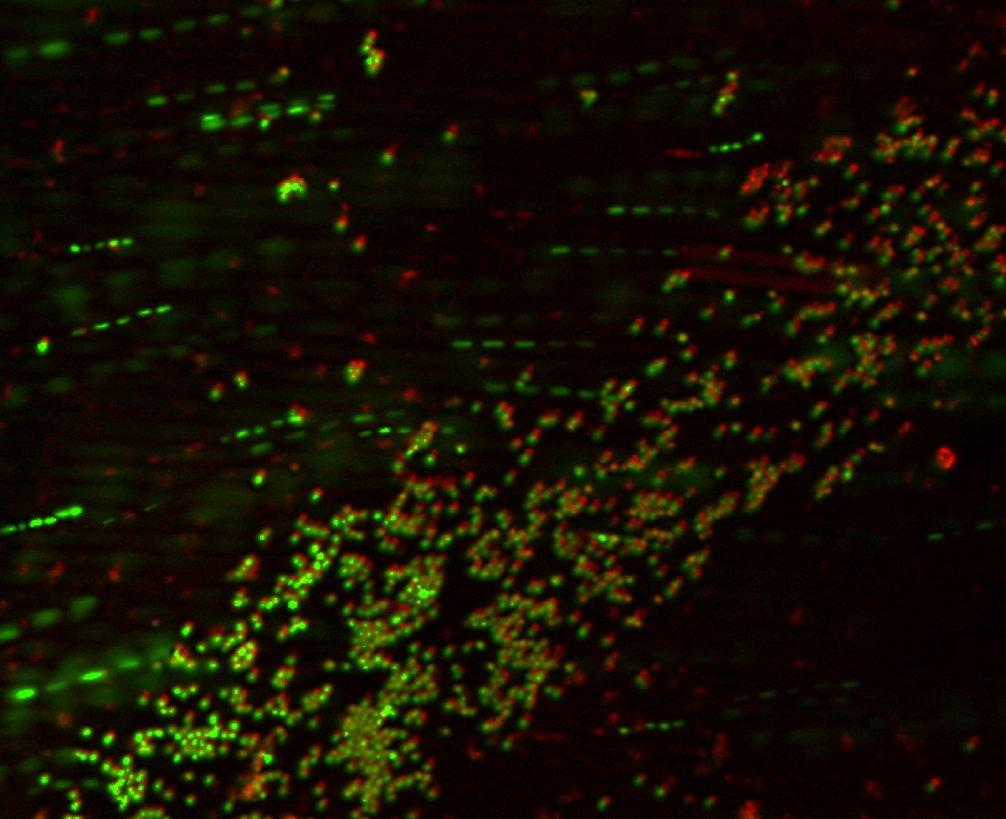
This image was taken during imaging of live bacteria with different dyes labelling parts of the cell. The image itself is blurred with the cells moving, and while not useful for my work, shows how beautiful the system can be even when it isn't behaving quite as we might hope.
Image: Rhia Stone | Related content: Cooper Group | Microscopy facility | Centre for Superbug Solutions
Conus textile having its lunch
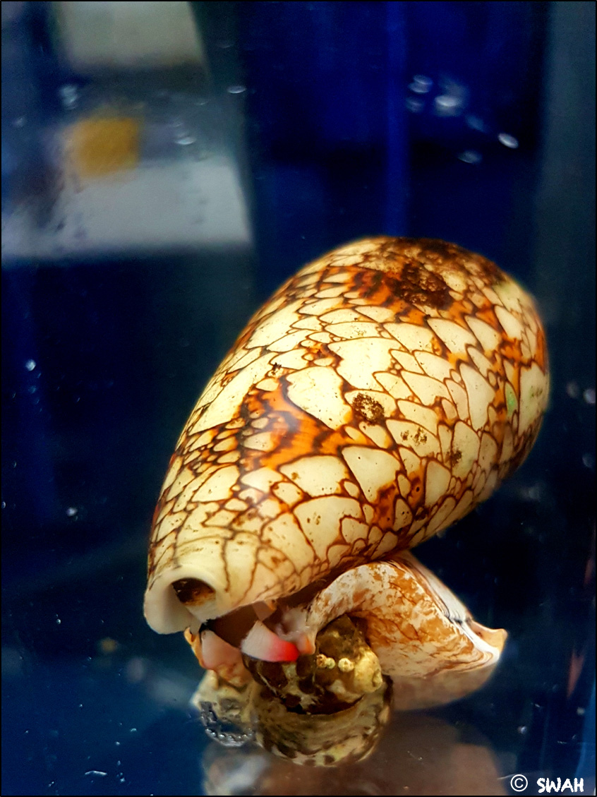
Cone snails use a complex venom to capture prey and deter predators. Their venom consist of mini proteins called conotoxins. These conotoxins can be developed as therapeutics to combat pain and neurodegenerative diseases. Therefore, what cone snails use to catch their lunch can be a treasure trove of therapeutics for us. This picture shows a mollusk-hunting conus textile predating on a Mulberry Whelk.
Image: Dr Himaya Siddhihalu Wickrama Hewage | Related content: Lewis Group | Centre for Pain Research
The vessels to the end of cardiovascular diseases
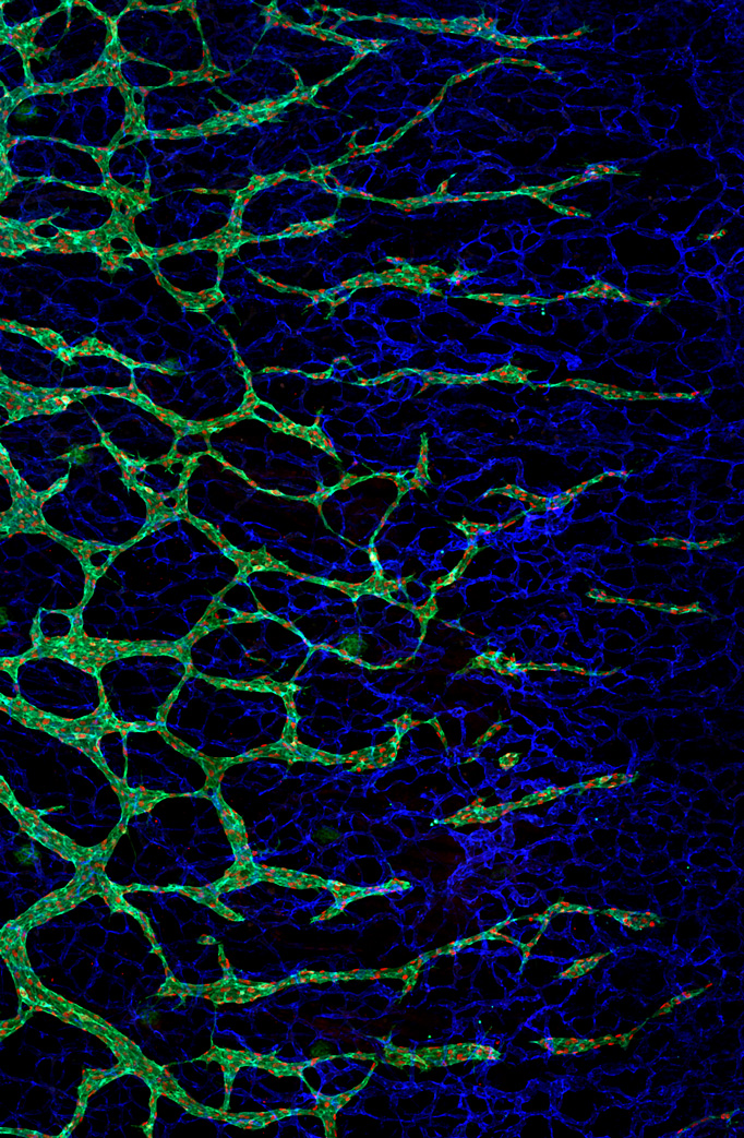
The image is the lymphatic (green) and blood (red) vasculatures. These vasculatures can be stained and imaged easily. We can better understand how different manipulations affect the development of cardiovascular network by studying these vasculatures. The knowledge of cardiovascular development will equip us to tackle cardiovascular diseases.
Image: Chui Ying (Tevin) Chau | Related content: Hogan Group | Microscopy facility | Centre for Cardiac and Vascular Biology
The endocardium and endothelium of a 24-hour-old zebrafish embryo
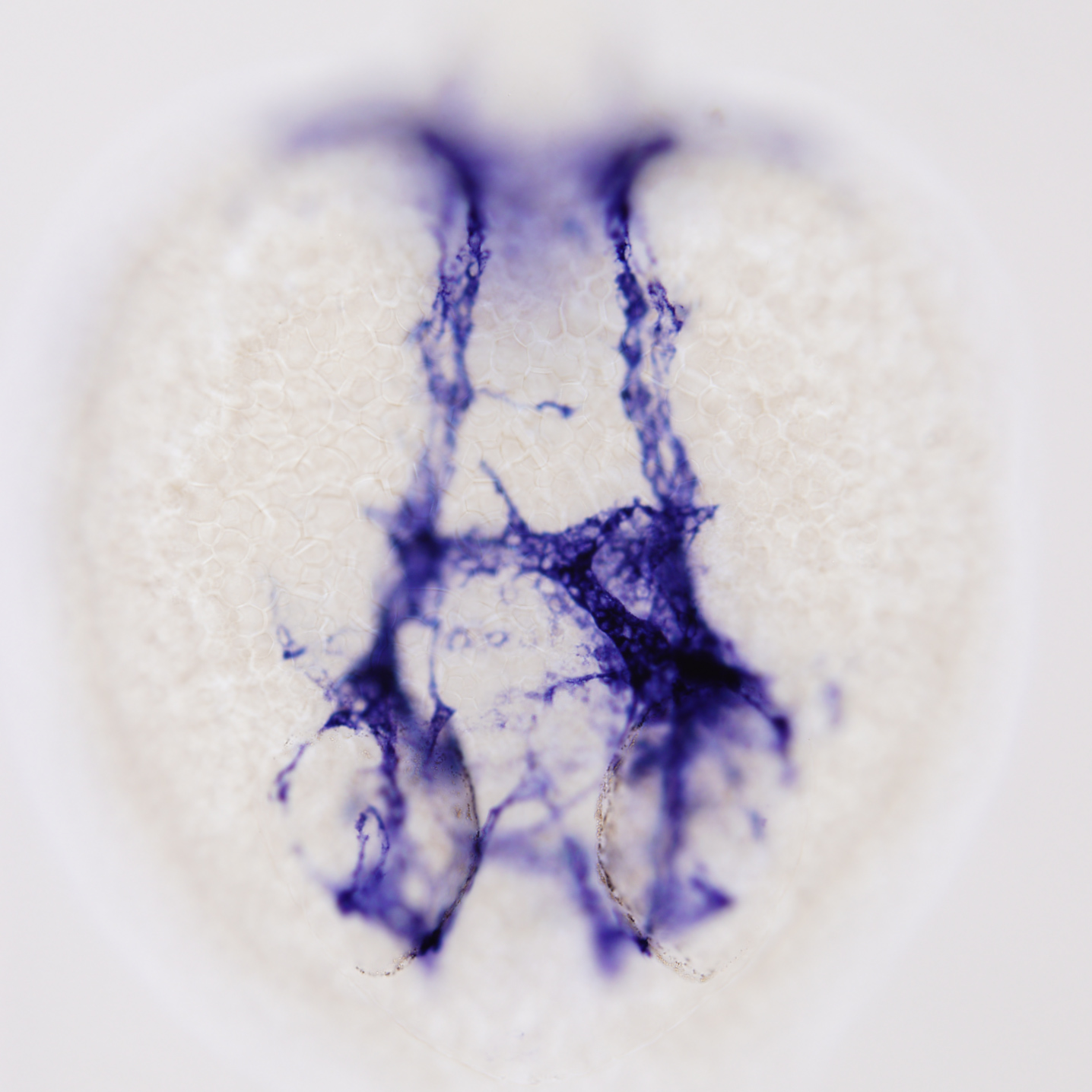
Image shows the expression of a marker throughout the circulatory system of a developing zebrafish embryo, dorsal view with the head to the bottom. This marker is expressed in blood vessels, but it is most highly expressed in the inner (endocardial) layer of the heart. The linear heart tube can be seen branching from the midline down towards the left eye of the embryo. Despite playing an important role in human health, the development of the endocardium is poorly understood. Using the expression of this marker in zebrafish as a tool to investigate endocardial development, we can follow the formation of the endocardium to discover the genes, signalling pathways and processes that lead to the formation of a functional heart.
Image: Sam Capon | Related content: Smith Group | Microscopy facility | Centre for Cardiac and Vascular Biology
Careful with that speck, Eugene
This is an image of an immune cell complex called an inflammasome, which is a major mediator of inflammatory and cell death responses to pathogens and cell stress signals. Understanding this process is critical in helping us discover new strategies to combat ever-increasing levels of infection in the community.
Image: Darren Brown | Related content: Stow Group | Microscopy facility | Centre for Inflammation and Disease Research
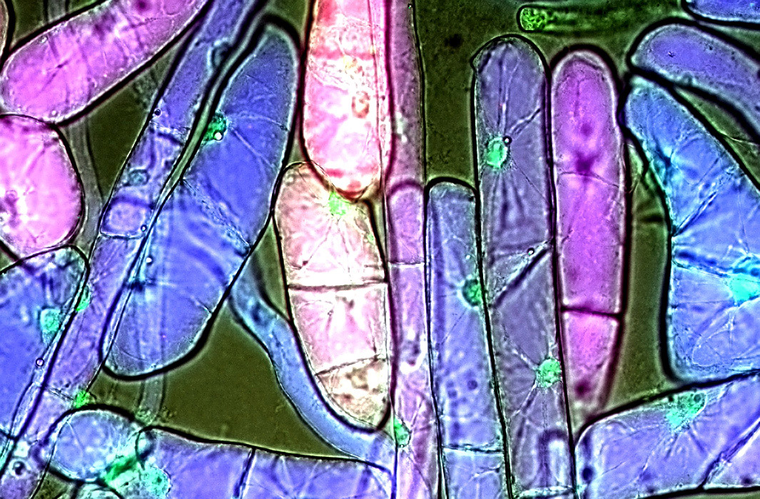
Ghost
African herb Oldenlandia affinis cells growing in suspension culture. To my knowledge, this is the only living suspension culture of O. affinis in the world. Adequate supply of pharmaceuticals is a major problem in a world of overpopulation and disparity. Biological production systems can help to inexpensively produce large quantities of engineered pharmaceuticals. This work focuses on the cultivation and analysis of Oldenlandia affinis cell suspension. The aim is molecular farming of therapeutic peptides grafted into a highly stable peptide-scaffold native to O. affinis. Stable cell suspension cultures were established and successfully upscaled to a 1L bioreactor. The morphology, viability, dry weight, packed cell volume, dissolved oxygen, pH and peptide expression were monitored.
Image: Benjamin Doffek | Related content: Craik Group | Clive and Vera Ramaciotti Facility for Producing Pharmaceuticals in Plants | Microscopy facility
The Neptune Valley
This is an image of an immune cell called a macrophage in the process of engulfing foreign particles. The image was composed from a raw data set taken from the 3View electron microscope at UQ. Our lab focuses on understanding the roles of key players in the inflammatory response. Our approaches are designed to understand the key host cell targets for the future development of new anti-microbial drugs and vaccines.
Image: Darren Brown | Related content: Stow Group | Centre for Inflammation and Disease Research
Spek-trom-i-ter
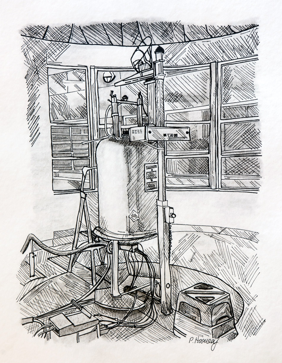
Nuclear magnetic resonance (NMR) spectroscopy exploits the magnetic properties of certain atomic nuclei and measures how these nuclei behave and interact when placed in a powerful magnetic field. It can be used to study the composition of biological or synthetic mixtures, the interaction of molecules, or the high-resolution structure of biological macromolecules such as proteins. This original, hand-drawn illustration depicts one of several NMR spectrometers within UQ’s Institute for Molecular Bioscience. Despite being our oldest spectrometer and one of the smallest, it is a faithful mainstay and continues to perform well with recent upgrades and industry-leading software.
Image: Dr Peta Harvey | Related content: Craik Group | Clive and Vera Ramaciotti Facility for Producing Pharmaceuticals in Plants | Nuclear Magnetic Resonance (NMR) Spectroscopy Facility
Crystal massacre
IMB researchers use a technique called X-ray crystallography to see the atomic detail of molecular machines. This involves growing crystals, picking them up, freezing them and shooting them with an X-ray beam then processing the data. This whole process can be challenging but this image depicts the difficulty associated with picking up microscopic crystals out of a 2 μL drop of liquid. It takes practice to perfect the technique and at the beginning, more often than not, your crystals don’t survive the process. This image is the result of one my very first attempts at picking up crystals. It took me so long the drop dried up under the heat of the microscope and what I was left with was a crystalline mess. This image serves as a reminder that failure is an inevitable part of the scientific process and is sometimes necessary for us to learn and grow.
Image: Emily Furlong | Related content: Collins Group | UQROCX Crystallisation Facility
Marley
This is an image of an immune cell called a macrophage during the process of inflammation. The image was composed from a raw data set taken from the 3View electron microscope at UQ, which was then assembled into 3D using 3D software. Our lab focuses on understanding the roles of key players in the inflammatory response and deciphering the key families of trafficking molecules involved. Our approaches are designed to understand the key host cell targets for the future development of new anti-microbial drugs and vaccines.
Image: Darren Brown | Related content: Stow Group | Centre for Inflammation and Disease Research
The cardiac cone
This image shows the expression of a marker of the developing zebrafish heart, dorsal view with the head to the top. This image shows a fluorescent, membrane-bound protein expressed in the beating muscle, cardiomyocytes, of the heart. At the stage shown the heart forms a cone shaped structure that will eventually extend telescopically into a linear heart tube. In zebrafish, the formation of the heart begins with the fusion of two progenitor populations into the cardiac cone. The heart begins to beat shortly after this. However, the heart still has to undergo significant developmental changes before it reaches its final form. The complexity of this process can lead to numerous health issues in humans at birth. Using zebrafish as a model of early heart development, we can uncover the processes that lead to the formation of a functional heart and gain insights into the treatment and prevention of congenital heart disease.
Image: Sam Capon | Related content: Smith Group | Microscopy facility | Centre for Cardiac and Vascular Biology
The Salmonella experience
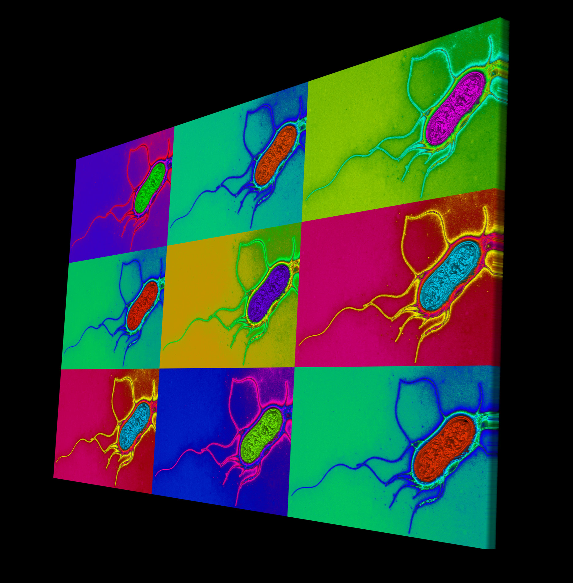
This is an image of Salmonella bacteria taken by a transmission electron microscope. Salmonella infection is a major cause of gastroenteritis. Our lab is trying to understand how bacteria and other external pathogens can cause disease and then try to identify and regulate the major players in our response to the resulting infection.
Image: Darren Brown | Related content: Stow Group | Centre for Inflammation and Disease Research
Migrating lymphatics 2
This image is showing a migrating lymphatic vessel (green stain shows the membrane of each cells forming the vessel, and blue stain shows nuclei of these cells) on top of the blood vessels (in red). This model expresses a red fluorescent protein in endothelial cells (cells forming the blood vessels). This genetic tool allows us to trace the endothelial cells (red) and all their progeny during development and will help to understand how lymphatics vessels form during development.
Image: Dr Cathy Pichol-Thievend | Related content: Francois Group | Microscopy facility | Centre for Cardiac and Vascular Biology
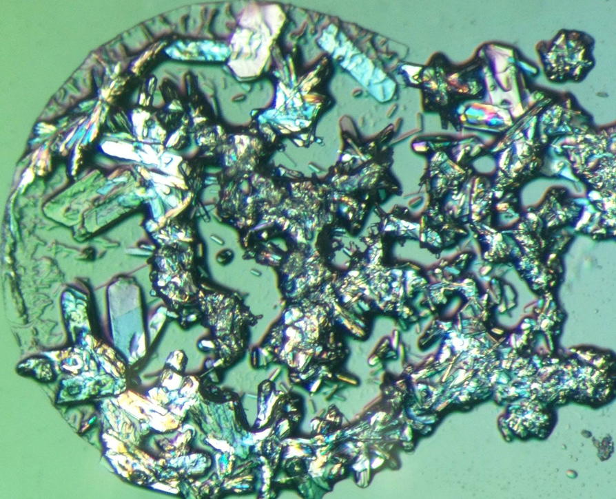
Cobalt chloride crystals
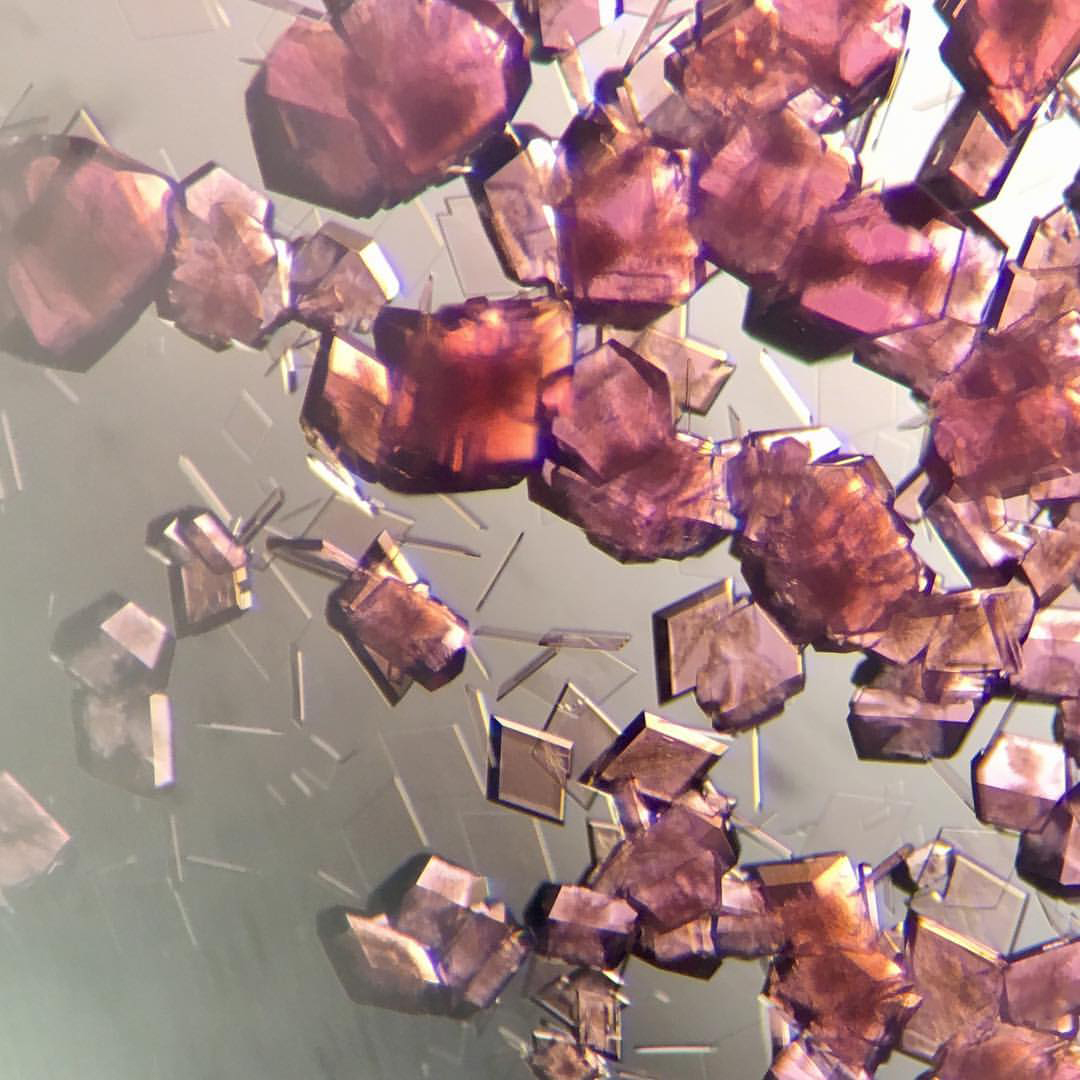
X-ray crystallography is a technique used by IMB researchers to study biological machines. This technique involves screening hundreds of crystal conditions to find one where the biological machine of interest will crystallise. Sometimes these screens will result in crystals of other things that aren't particularly useful to the scientist. This photograph is of cobalt chloride crystals that were obtained in a crystallisation experiment.
Image: Emily Furlong | Related content: Collins Group | UQROCX Crystallisation Facility
Great science comes with great patience
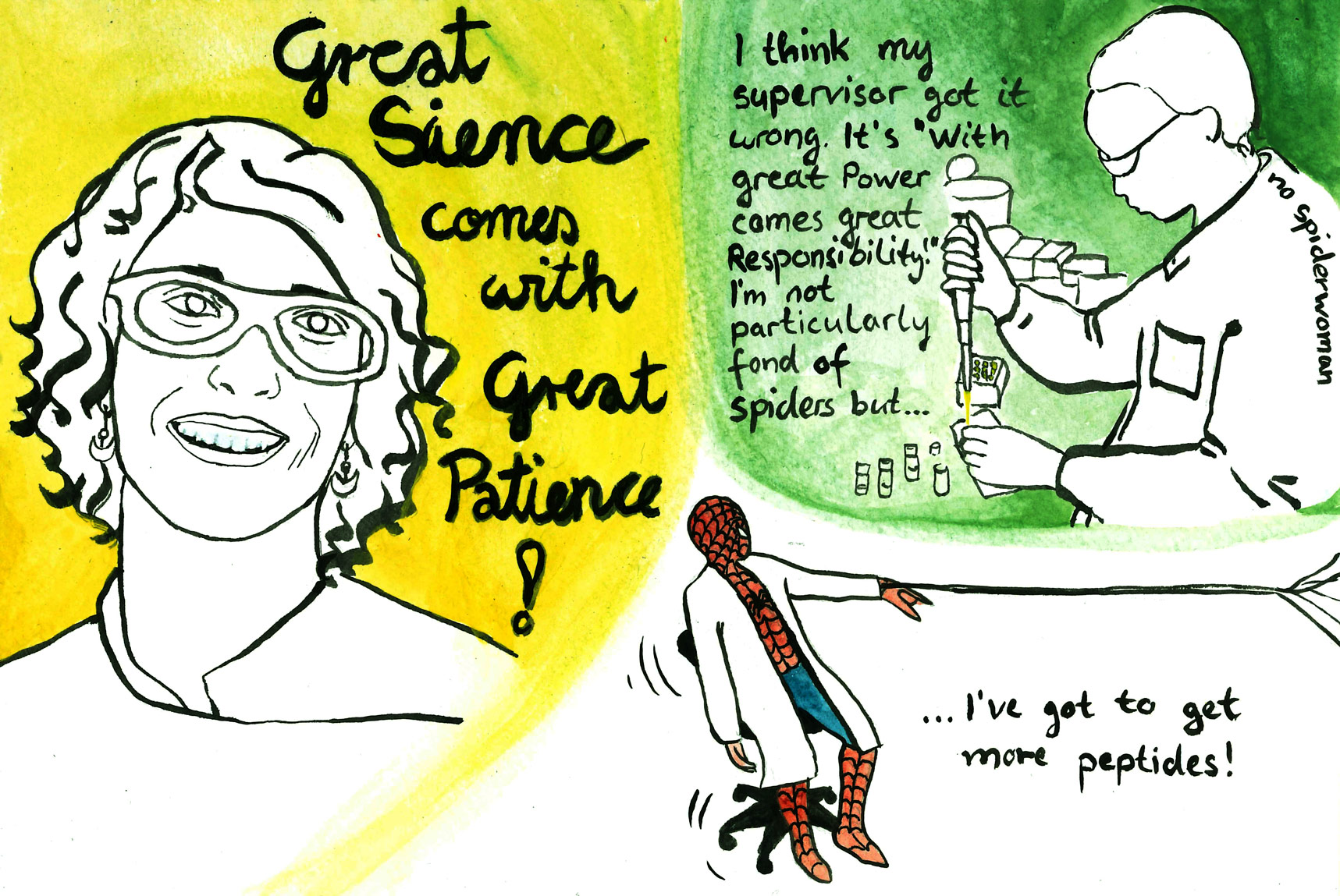
This quote of my supervisor reminded me strongly of Spiderman and it just happens to be that one of her other students is actually working on converting peptides from the venom of tarantula spiders into potential pain therapeutics.
Image: Felicitas Vernen | Related content: Craik Group
The gathering
African herb Oldenlandia affinis cells growing in suspension culture. To my knowledge, this is the only living suspension culture of O. affinis in the world. Adequate supply of pharmaceuticals is a major problem in a world of overpopulation and disparity. Biological production systems can help to inexpensively produce large quantities of engineered pharmaceuticals. This work focuses on the cultivation and analysis of Oldenlandia affinis cell suspension. The aim is molecular farming of therapeutic peptides grafted into a highly stable peptide-scaffold native to O. affinis. Stable cell suspension cultures were established and successfully upscaled to a 1L bioreactor. The morphology, viability, dry weight, packed cell volume, dissolved oxygen, pH and peptide expression were monitored.
Image: Benjamin Doffek | Related content: Craik Group | Clive and Vera Ramaciotti Facility for Producing Pharmaceuticals in Plants | Microscopy facility
Jimi's castle
This is an image of an immune cell called a macrophage in the process of engulfing foreign particles. The image was composed from a raw data set taken from the 3View electron microscope at UQ. Our lab focuses on understanding the roles of key players in the inflammatory response and deciphering the key families of trafficking molecules involved. Our approaches are designed to understand the key host cell targets for the future development of new anti-microbial drugs and vaccines.
Image: Darren Brown | Related content: Stow Group | Centre for Inflammation and Disease Research
How to break a chemist's heart
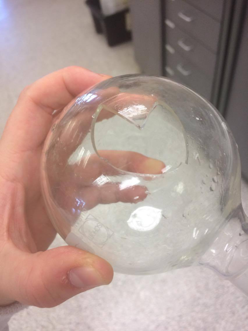
When flasks break just right! This is a light-hearted image of when my round-bottom flask broke in a nice heart shape. No product was lost in the making of this image.
Image: Rhia Stone | Related content: Cooper Group
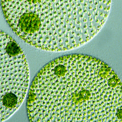
Got algae?
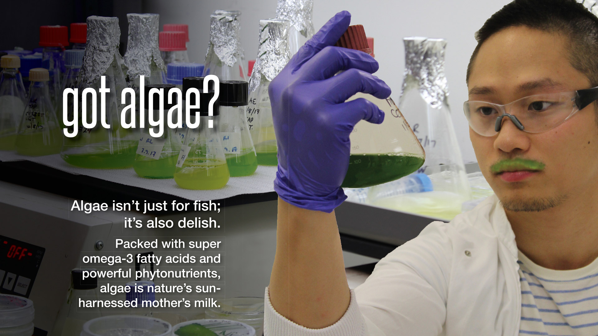
Got milk? Well, we’ve got something better. As demands for food and freshwater increase with our rapidly expanding population, these tiny microalgae can use sunlight, non-arable land, and CO2 to produce new foods rich in omega-3 fatty acids, proteins and phytonutrients. The future looks green!
In collaboration with Jennifer Yarnold and Dake Xiong and inspired by the “Got milk?” commercials.
Image: Felicitas Vernen | Related content: Craik Group | Hankamer Group | Centre for Solar Biotechnology