Objective Selection
Introduction
Not all objectives are made equal! Objectives for microscopes contain lots of very small and delicate lens to both magnify the image, as well as to perform a number of corrections (spherical and chromatic). Two main characterizations of objectives are the magnification and the numerical aperture.
There are different 'grades' of objectives which describe how many colours they are axially corrected for, and how many colours they are spherically corrected for.
Achromats (Axial 2 channels, Spherical 1 channel)
Fluorites (Axial 2-4 channels, Spherical 2-4 channels)
Apochromats (Axial 4-5 channels, Spherical 3-4 channels)
Some objectives are also "Plan-" objectives, these are flat field corrected, so the image appears flat to both the eyepieces and the detector.
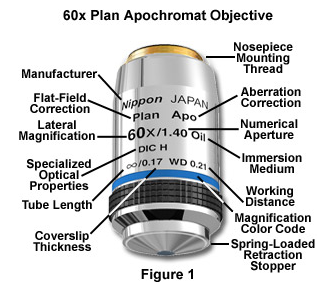
Key markings on objectives.
Source Mortimer Abramowitz & Michael Davidson
Magnification & Numerical Aperture
Numerical Aperture
The resolving power of the objective is determined by the numerical aperture (not magnification). Numerical aperture also determines the light collecting ability of the objective, the higher the NA the more light the objective can collect and is determined by the function (nSinq, Where n = immersion media refractive index, and Sinq = angle of the cone of illumination).
For reference the NA of common immersions are:
Air = 1.0
Water = 1.33
Oil = 1.515
Microscopy: Objectives and Eyepieces (Stephen Ross)
Numerical Aperture
Brightness
Image brightness is directly proportional to the objective NA and inversely proportional to magnification. Therefore if you had two objectives of the same NA but differed in magnification, for examples the following;
Nikon 60x 1.4NA = 10.7 Arbritrary Brightness units
Nikon 100x 1.4NA = 3.8 Arbritrary Brightness units
The 60x 1.4NA objective is ~2.8x brighter than the 100x and both objectives have the same resolution!
The terms F(trans) and F(epi) refer to the light-gathering power of an objective and were calculated according to the following equations:
F(trans) = 104 × NA2/M2
F(epi) = 104 × (NA2/M)2
Where F(trans) refers to image brightness of a Brightfield illuminated sample, whereas F(epi) refers to the image brightness of a fluorescence or reflected light illuminated sample.
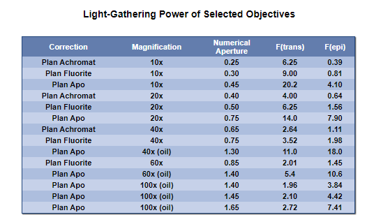
Table of example objectives and their brightness for both Transmitted and epifluorescence modalities.
Source: Kenneth Spring, Ernst Keller, Michael Davidson, Anatomy of the Microscope.
In theory, the intensity of illumination depends on the square of the condenser numerical aperture and the square of the demagnification of the light source image (in effect, the field diaphragm image becomes brighter as it is made smaller, according to the square law). The result is that brightness of the specimen image is directly proportional to the square of the objective numerical aperture as it reaches the eyepiece (or camera system), and also inversely proportional to the objective magnification. Therefore, when examining specimens in transmitted light, changing the objective without altering the condenser affects image brightness in response to changes in numerical aperture and magnification.
Image Brightness calculator: Objective Relative Brightness Calculator.xlsx
This excel spreadsheet has two worksheets that let you compare different objectives to determine their relative brightness
Special Objectives
Phase
https://www.microscopyu.com/microscopy-basics/specialized-microscope-objectives
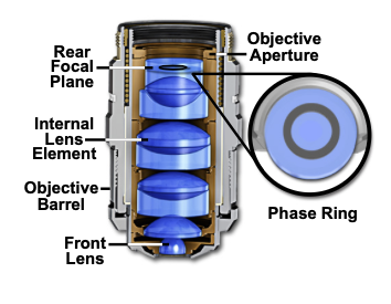
A special objective is required that is fitted with a darkened circular ring or groove (phase plate) fitted into the glass near the rear focal plane of the objective as illustrated in Figure 1. In addition, the condenser must also be modified with special annular openings suited to a particular magnification and objective. Phase contrast objectives are segregated into a number of categories depending upon the construction and neutral density of internal phase rings:
- DL (Dark Low) - DL objectives produce a dark image outline on a light gray background. These objectives are designed to furnish the strongest dark contrast in specimens having major differences in refractive indices. The DL phase contrast objective is the most popular style for examination of cells and other semi-transparent living material and is especially suited for photomicrography and digital imaging.
- DLL (Dark Low Low) - Similar to the DL objective, the DLL series allows better images in brightfield and is often used as a "universal" objective in microscope systems that utilize multiple illumination modes such as fluorescence, DIC, brightfield, and darkfield.
- ADL (Apodized Dark Low) - Recently introduced by Nikon, the apodized phase contrast ADL objectives contain a secondary neutral density ring on either side of the phase ring. Addition of the secondary rings assists in reducing unwanted "halo" effects often associated with imaging in phase contrast microscopy.
- DM (Dark Medium) - DM objectives produce a dark image outline on a medium gray background. These objectives are designed to be used for high image contrast with specimens having small phase differences, such as fine fibers, granules, and particles.
- BM (Bright Medium) - Often referred to as negative phase contrast, BM objectives produce a bright image outline on a medium gray background. BM objectives are ideal for visual examination of bacterial flagella, fibrin bundles, minute globules, and blood cell counting.
In order to allow the microscopist to quickly identify phase contrast objectives, many manufacturers inscribe important specifications, such as the magnification, numerical aperture, tube length correction, etc., on the outer barrel in green letters. This serves to differentiate phase contrast objectives from ordinary brightfield, polarized, DIC, and fluorescence objectives which either use an alternative color code or the standard black lettering.
Correction Collars
Correction collars are generally used to correct for spherical aberration due to variations in coverslip thickness, temperature, wavelength or for the differing refractive indices of different immersion media.
Most microscope objectives are designed to be used with a cover glass that has a standard thickness of 0.17 millimeters and a refractive index of 1.515, which is satisfactory when the objective numerical aperture is 0.4 or less. However, when using high numerical aperture dry objectives (numerical aperture of 0.8 or greater), cover glass thickness variations of only a few micrometers result in dramatic image degradation due to aberration, which grows worse with increasing cover glass thickness. To compensate for this error, the more highly corrected objectives are equipped with a correction collar to allow adjustment of the central lens group position to coincide with fluctuations in cover glass thickness.
https://www.microscopyu.com/tutorials/adjustment-of-objective-correction-collars
Microscopy: Correcting for Spherical Aberration with a Correction Collar (Stephen Ross)