Dragonfly Spinning Disc
Confocal with TIRF
.
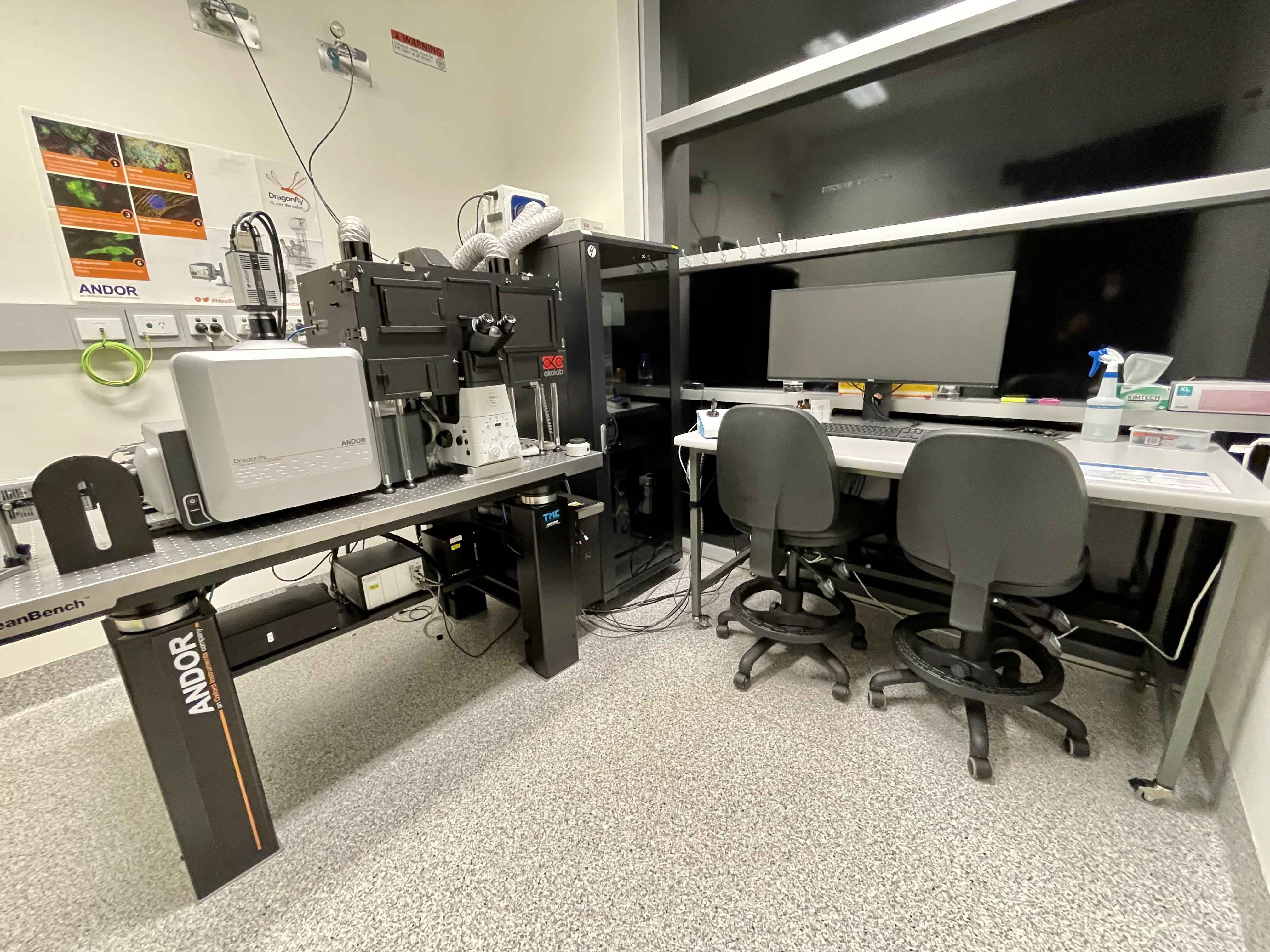 Room 6.025b - Nikon Ti2 Inverted Microscope Stand with Andor Dragonfly Spinning Disc Scanhead
Inverted spinning disc laser confocal microscope, suited for fast live imaging, 3-dimensional multi-position and large scale tiling. Also has TIRF functionality and incubation.
Features Include:
- Fully motorised X-Y-Z stage
- 500 um Piezo Z-drive for rapid Z-stacks
- CO2 and temperature incubation for live cells and tissue
- Tiled imaging
- Multi-position imaging
- TIRF imaging
- Routine time-lapse and ultra-fast burst mode imaging
- Laser Widefield mode
- Nikon Perfect Focus 4
Transmitted Light: CoolLED White Light LED
Reflected Light: Nikon Intensilight 100W White Light Lamp
Installed in March of 2018 is the Andor Dragonfly incorporates a Spinning Disc Confocal allowing researchers to image 3D volumes at greater speed and over larger areas than ever before.
.
Condensor
| Name | N.A. | W.D. mm | Position 1 | Position 2 | Position 3 | Position 4 | Position 5 | Position 6 |
| Universal LWD | - | - | ND | - | - | DIC I | DIC II | C |
Objectives
| Position | Objective | Mag | N.A. | Imm | W.D. (mm) | Type | R.I. | XY Res (nm) | Arbitrary Brightness |
|---|---|---|---|---|---|---|---|---|---|
| 1 | Plan Apo | 4x | 0.2 | Dry | 20 | - | 1.00 | 1488 | 1 |
| 2 | Plan Apo λ | 10x | 0.45 | Dry | 4.0 | DIC I | 1.00 | 662 | 4 |
| 3 | Plan Apo VC | 20x | 0.75 | Dry | 1.0 | DIC II | 1.00 | 397 | 8 |
| 4 | Plan Apo λ | 40x | 0.95 | Dry | 0.16-0.25 | DIC II | 1.00 | 313 | 5 |
| 5 | Apo λ LS | 40x | 1.15 | Water | 0.59-0.61 | DIC II | 1.33 | 259 | 11 |
| 6 |
Plan Apo λ |
60x |
1.4 |
Oil |
0.13 |
Ph3 DM |
1.515 |
213 | 11 |
*25x Silicone Oil objective is also available (1.05 N.A.)
*100x TIRF Oil objective is also available (1.49 N.A.)
Laser Lines Available
405, 445, 488, 514, 561 and 637nm
Eyepiece Fluorescent Filter Sets
| Position | Name | Excitation | Dichroic | Emission | Suitable Dyes |
|---|---|---|---|---|---|
| 1 | DAPI | 375/28 | 415 | 460/60 | ECFP, Cerulean |
| 2 | FITC | 480/30 | 505 | 535/46 | EGFP, Alexa Fluor 488 |
| 3 | TRITC | 540/26 | 565 | 605/56 | EYFP, Citrine |
| 4 | - | - | - | - | - |
| 5 | - | - | - | - | - |
| 6 |
- |
- | - | - | - |
Confocal Fluorescence Dichroic Sets
|
First Dichroic Set |
Second Dichroic Set | Third Dichroic Set |
|---|---|---|
|
405-13 |
405-13 | 405-13 |
| 488-13 | 445-13 | 488-13 |
| 561-6 | 514-14 | 561-6 |
| 640-16 | 640-16 | 685-13 |
Simulataneous Acqisition Image Splitter
| Image Spliiter |
|---|
| 100% pass |
| 500 nm Long Pass |
| 565 nm Long Pass |
| 640 nm Long Pass |
Confocal Emission Filter Wheel Options
| Position | Camera 1 | Camera 2 |
|---|---|---|
| 1) | 620-60 | TR-442-514-561 |
| 2) | 600-50 | 600-50 |
| 3) | 542-27 | 525-50 |
| 4) | 525-50 | 483-32 |
| 5) | 480-70 | 450-50 |
| 6) | 450-50 | TR-POLR-DIC-FWL |
| 7) | TR-POL-DIC-FWL | Blocked |
| 8) | 700-75 | Blocked |
Computer
Dell Precision 7910 PC - 3.5GHz Xeon Processor - 96GB RAM - 8GB nVidia Quadro M4000 GPU
4TB Raid Array
30" LCD Monitor
Software
Fusion and Imaris
Accessories
Incubation chamber with CO2 and Temperature control
Single Microscope Slide + 2x35 mm dish insert
Contacts
Dr James Springfield
Microscopy Facility Manager
+61 7 334 62390
j.springfield@imb.uq.edu.au
Dr Nicholas Condon
CZI Imaging Scientist
+61 7 334 62042
n.condon@imb.uq.edu.au
Dr Mahdie Mollazade
Microscopy Officer
+61 7 334 62042
m.mollazade@uq.edu.au
Mailing address
Advanced Microscopy Facility
Institute for Molecular Bioscience
Level 6N, 306 Carmody Road,
Building 80
University of Queensland
4072, St Lucia,
Queensland, Australia