Spinning Disc Confocal
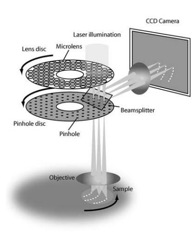
Spinning Disc Confocal (SDC) microscopes combine the optical sectioning benefit of a traditional single point scanning confocal microscope with the temporal benefits of a camera based widefield microscope. This is achieved through parallelization of the confocal pinhole, by spinning a disc with many pinholes over a large area, a single camera exposure can be used to capture the optical section, rather than building up the image pixel by pixel.
Modern spinning disc confocal systems have twin discs, one containing the confocal pinholes, and one containing micro-lenses. Illumination light first passes through the microlens disc where it is focused through to their matched pinholes. Yokogawa first introduced the microlens array to improve illumination efficiencies, and is now industry standard. Emission light from the sample then returns through the pinhole disc for optical sectioning before being directed towards the detection camera via a dichroic mirror while also preventing any laser back-scatter.
In addition to the speed benefits, camera-based detectors typically have much higher quantum efficiencies compared to photon-multiplier tubes (PMTs) found in traditional confocal microscopes.
Source: Teledyne Photometrics
Contacts
Dr James Springfield
Microscopy Facility Manager
+61 7 334 62390
j.springfield@imb.uq.edu.au
Dr Nicholas Condon
CZI Imaging Scientist
+61 7 334 62042
n.condon@imb.uq.edu.au
Dr Mahdie Mollazade
Microscopy Officer
+61 7 334 62042
m.mollazade@uq.edu.au
Mailing address
Advanced Microscopy Facility
Institute for Molecular Bioscience
Level 6N, 306 Carmody Road,
Building 80
University of Queensland
4072, St Lucia,
Queensland, Australia