Image Deconvolution
Background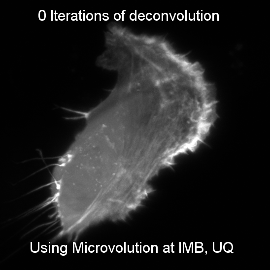
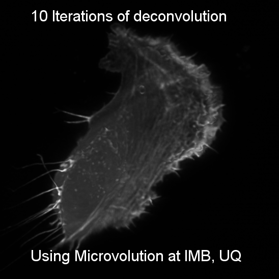
Wide-field microscopy involves illumination of the whole sample, and relies on the optical characteristics of the imaging system to capture a single focal plane. The limitation of these kinds of imaging systems is that often, out of focus light (from differing sample planes) are captured in your images, resulting in image-blur. This image blur will lower your signal to noise, which is the contrast of your images. One way to overcome this is by using specialized optics with very narrow depths of field (High NA objectives) coupled with computational algorithms that can correct for out of focus light, this process is called deconvolution.
The light coming from your sample scatters (diffraction) in all directions when illuminated, and this can be affected by coverslip thickness, sample mounting medium and the many components of the microscope between your sample and the camera. The way that the light diffracts can be characterized by the point spread function (PSF) whereby a single point of light of known size (eg fluorescent bead) is captured in xyz and shows the scattering behaviour of light in all dimensions for that particular optical system. The PSF is then used to reverse the effects of out of focus light by correcting for these known distortions in all dimensions.
IMB Deconvolution Presentation
External Deconvolution Links
- iBiology Video
- Micro Course Video - PSF
- A Workingperson's Guide to Deconvolution in Light Microscopy – BioFeature Article
- To 5D and Beyond: Quantitative Fluorescence microscopy in the Postgenomic Era – Traffic Article
Deconvolution software available at IMB
- Microvolution (Vendor Website)
- Huygens (Vendor Website)
- Leica Lightning (Vendor Website)
- Andor Fusion (Vendor Website)
- Softworx (Only available on Deltavision Microscope)
Microvolution
Microvolution is a multi-GPU compatible deconvolution licensed package that runs within FIJI for you convenience. We have purchased licenses for Microvolution to run on the virtual computers as well as on Wiener.
Availability
- Cupid VM
- Hercules VM
- Lucifer VM
- Saturn VM
- Wiener (requires specific access, email microscopes@imb.uq.edu.au for more information)
Guides
- Microvolution manual - IJ.pdf
- Generated PSF Deconvolution Procedure.pdf
- Measured PSF Deconvolution Procedure.pdf
- LLSM Measured PSF Deconvolution Procedure.pdf
Huygens
Huygens deconvolution software available at IMB came included with the purchase of the Leica SP8 Confocal prior to the roll out of Leica's own Lightning deconvolution package. It can be used for confocal and widefield deconvolution and includes the necessary packages for STED deconvolution.
Availability
- Saturn VM
Guides
Note: This guide was produced by Rumelo Amor from QBI and is based on their platform of using X-11 for remote access. The IMB version runs in Windows on Saturn only. Relevant information begins from about page 9.
Leica Lightning
Lightning is Leica's deconvolution package with is installed on the SP8 confocal. Where Leica's product differs from other deconvolution products is the number of cycles performed on the image is pixel dependant, meaning one area of an image may have 20 cycles performed, while another may only have 5. This method is very novel and has not been implemented by any other vendor. The results are reproducible, as the software isn't using machine learning, and with the fast parallel GPU processing the deconvolution is performed nearly in real time.
The optimum parameters for the subsequent deconvolution are determined for each associated volume segment around each pixel. This adaptive process of correlating the deconvolution parameter space with the local image properties enables a fully automated and voxel-accurate information recovery.
The deconvolution parameters are linked to:
1. Microscopic hardware, imaging and experimental setup: Excitation/emission wave lengths, objective lens, resolution, sample substrate, immersion/embedding media, etc.
2. Image characteristics: Background, SNR, regularization (the correct interpretation of background or noise by the algorithm distinct from information-carrying signal), number of iterations, etc which informs a pixel-wise 'decision map'
At present Leica does not release the 'decision map' image, however it is rumoured to be coming soon.
Traditional deconvolution methods use a 'best guess', global approach, through which the maximum possible trade-off in the creation of the global deconvolution parameter space is achieved. The disadvantage of this approach is that it does not take inhomogeneities of the image into account.
Availability
- SP8 Confocal Microscope only