Lattice Lightsheet Microscope
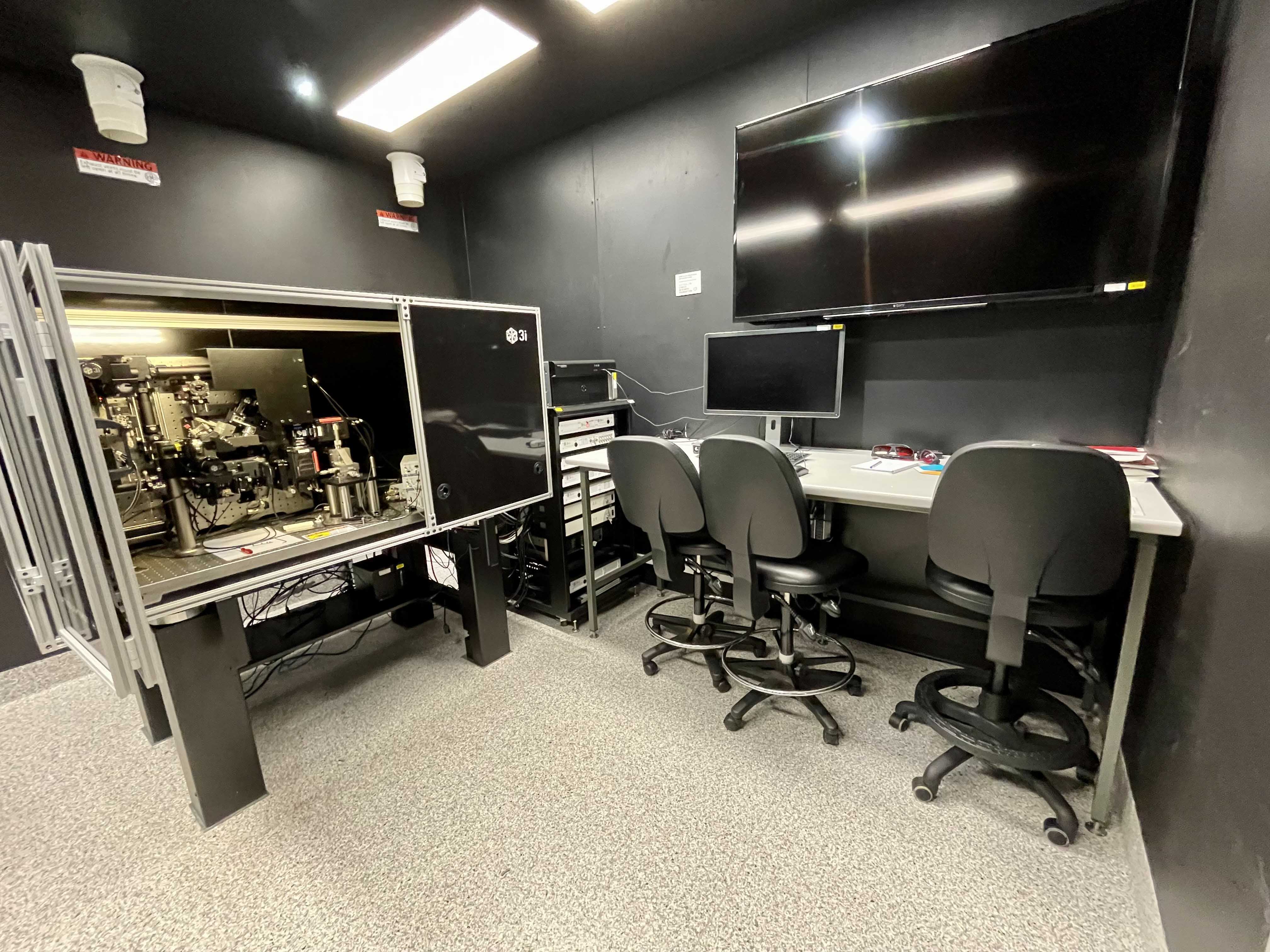
Room 6.019b
The Lattice Lightsheet microscope is an advanced live cell microscope developed by Nobel Prize winner Eric Betzig in 2014. This microscope is extremely gentle on cells allowing for very long acquisitions, as well as being super-fast, (capturing full 3D volumes in under a second).
Highlights
- Very High spatial resolution (230 nm, 230 nm, 300 nm, xyz)
- Very High Temporal resolution (100 slices / second)
- Very low photo-toxicity
- Opportunity to generate very rare data (very few working systems world wide)
- 4 Individual lasers (440 nm (CFP), 488 nm (GFP), 560 nm (RFP), 640 nm (Far Red))
Challenges
- Massive data sets are generated by this system (terabytes)
- Visualisation and analysis software solutions are not equipped for large volumes of LLSM data, instead MIPS are generated for indexing of files, and finding sections of movies for further analysis. Large collaborations are underway to develop tools to fix this.
- This system is not a ‘turnkey solution’ like our Zeiss systems, therefore the software is not as refined, and often many alignments are required to optimise the imaging output.
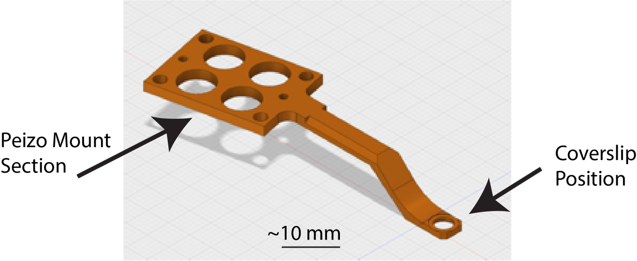
Sample Preparation
- This microscope takes 5 mm coverslips (thickness is not important) mounted on the holder shown below.
- The stage can be moved within a 3 x 3 mm region from the centre of the coverslip
- Samples must be adhered to the coverslip (for non-single cell imaging please consult with facility staff)
- Samples are imaged with water dipping lens’ with your sample in a 8 mL water bath (take this into account if your require stimulants/drugs in your imaging media)
- The stage/water-bath is heated, however CO2 is not available while imaging.
- The LLSM room has an incubator set to 37 oC 5% CO2.
To submit a proposal for Lattice Lightsheet Imaging, please use this form.
“For the first time we will be able to see the behaviour of cells and organisms in real time at high resolution. Previous microscopes damage the cells, but the Lattice Light Sheet does not, so we will also be able to watch generations of cells over several days giving comprehensive insights into their behaviour.
“We will observe the impact of drugs on cells and disease-causing mutations on cells very quickly and accurately. This technology will greatly enhance our understanding of cells normal behaviour and our understanding of diseases like cancer and inflammation. It will also accelerate research into treatments.” Professor Jenny Stow, Leader The Stow Group - Protein Trafficking and Inflammation.
Contacts
Dr James Springfield
Microscopy Facility Manager
+61 7 334 62390
j.springfield@imb.uq.edu.au
Dr Nicholas Condon
CZI Imaging Scientist
+61 7 334 62042
n.condon@imb.uq.edu.au
Dr Mahdie Mollazade
Microscopy Officer
+61 7 334 62042
m.mollazade@uq.edu.au
Mailing address
Advanced Microscopy Facility
Institute for Molecular Bioscience
Level 6N, 306 Carmody Road,
Building 80
University of Queensland
4072, St Lucia,
Queensland, Australia