Deconvolution
Wide-field microscopy involves illumination of the whole sample, and relies on the optical characteristics of the imaging system to capture a single focal plane. The limitation of these kinds of imaging systems is that often, out of focus light (from differing sample planes) are captured in your images, resulting in image-blur. This image blur will lower your signal to noise, which is the contrast of your images. One way to overcome this is by using specialised optics with very narrow depths of field (High NA objectives) coupled with computational algorithms that can correct for out of focus light, this process is called deconvolution.
The fluorescent signal coming from your sample scatters (diffraction) in all directions when illuminated. The image blur stems from the many optical components that the light has passed through in the microscope between your sample and the camera. However, the light scatter occurs in a predictable manner based on some optical parameters that you can take note of during image acquistion:
- wavelength
- objective and its numerical aperture (NA)
- refraction index of the immersion media
- coverslip thickness
And others that you cannot control such as the adjustment of the system and other optical parameters.
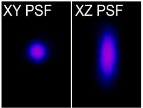
The accumulated result of all these factors on a single point object is described as a point spread function (PSF). Therefore, if a single point of light of known size (fluorescent bead) is captured in xyz, then the scattering behaviour of light in all dimensions for that particular optical configuration can be characterised by the PSF. The PSF is then used to reverse the effects of out of focus light by correcting for these known distortions in all dimensions, the process is called deconvolution.
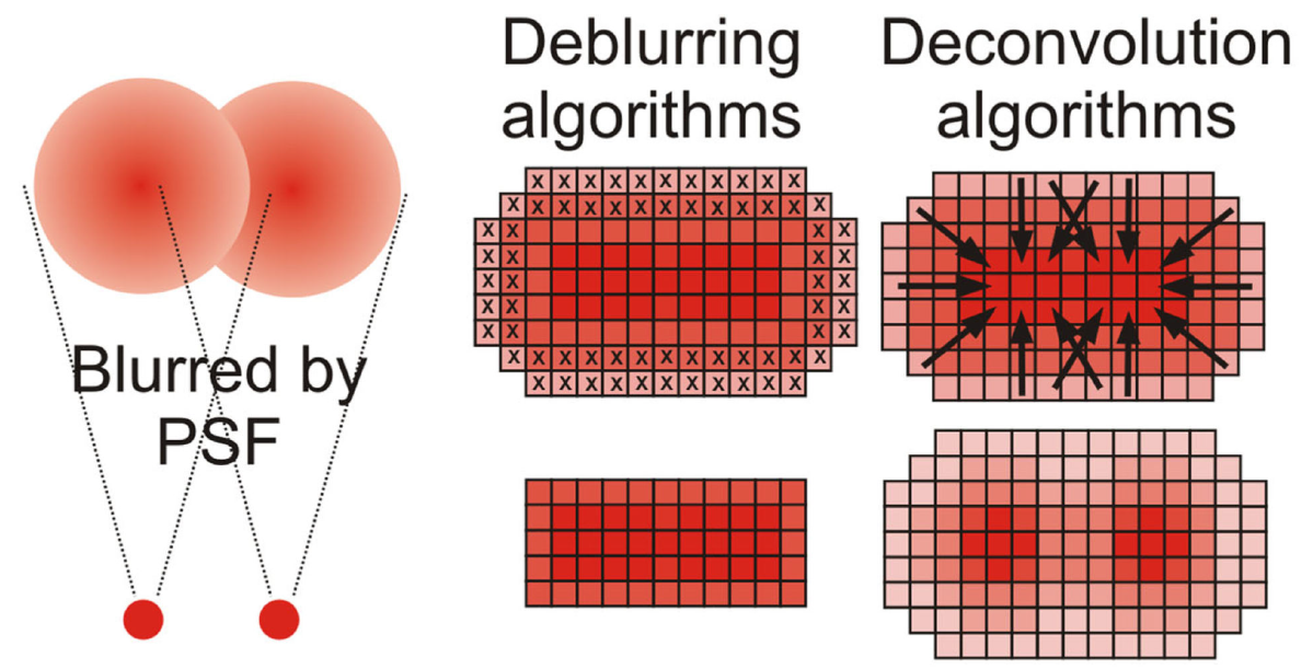
Verdaasdonk, J.S. 2014
Deconvolution is an iterative, post-processing method which is performed on your images and described in the image analysis section.
In order to successfully deconvolve your images, you must ensure they were captured with the correct Nyquist rates as this cannot be fixed afterwards.
Deconvolution on Leica SP8
Lightning Settings
In this dialog, enter the required data for the specimen and define the image optimization strategy.
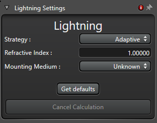
- Mounting Medium: Here, you select the embedding medium.
- Refractive Index: This displays the refractive index of the selected embedding medium.
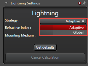
- Strategy:
- Adaptive: On the basis of the SNR, the settings for optimum image quality are determined automatically.
- Global: This strategy is recommended as default for all images.
- Get defaults: Resets the settings to the default values.
- Cancel Calculation: Stops calculation of the automatic image optimization. This button is not enabled until the image acquisition has been stopped, while the calculation is still in progress. This function enables you to cancel the calculation process prematurely, for example, if you started a long image acquisition (such as a xyzt series) and realize that the settings are not optimal and you would like to restart the series with adjusted settings. Then you do not have to wait until the calculation is done. The cancellation occurs when the currently calculated series is done.
Other resources: