Image Analysis
The Imaging facility currently has 4x dedicated, physical workstation computers in Room 6.055 as well as a number of virtualised machines available for image processing and visualisation of your microscopy data.
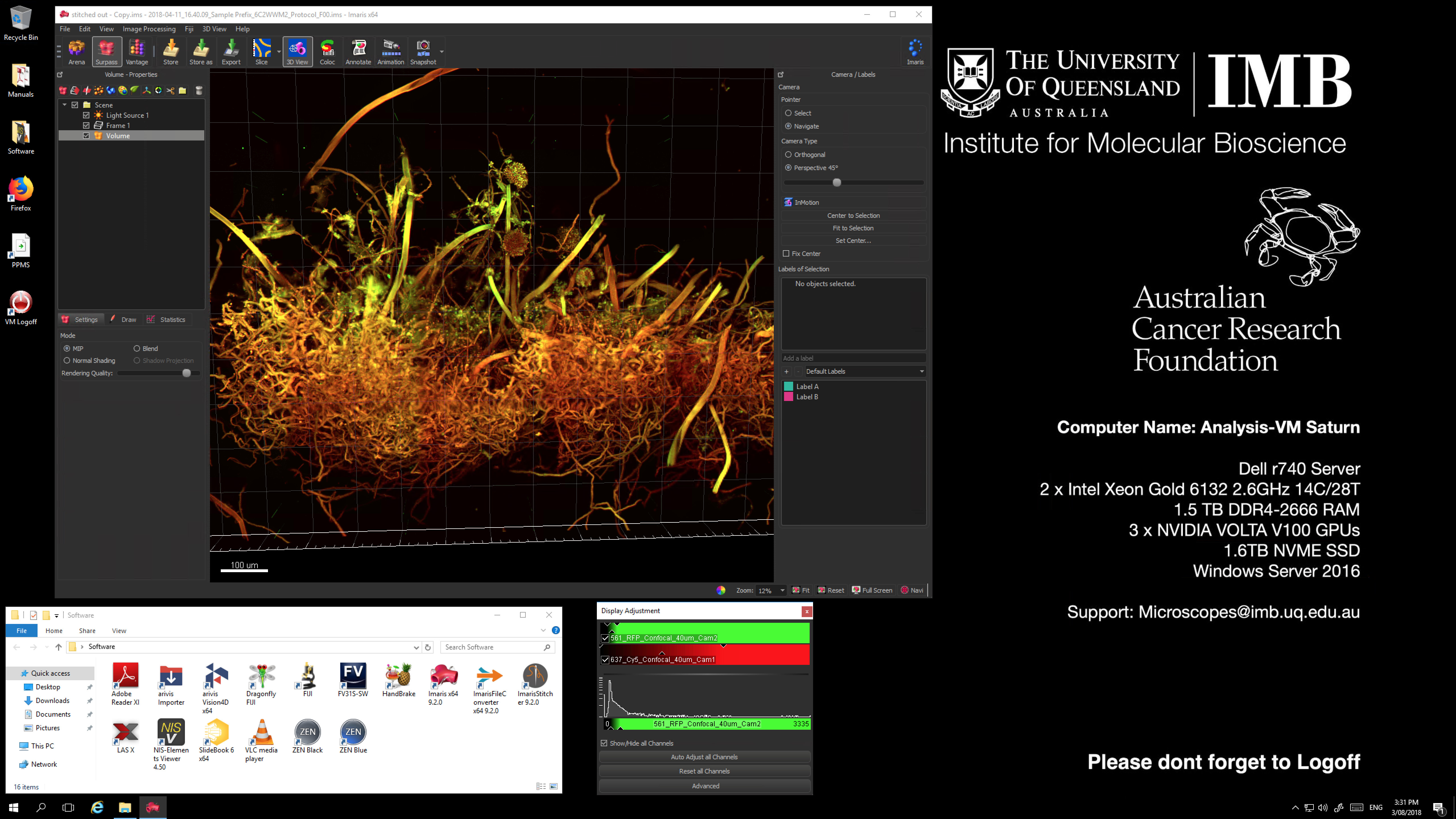
Analysis virtual machines are high-powered, graphics accelerated computers available to all users of the imaging facility to access, either onsite at the Queensland Bioscience Precinct, or remotely via VPN. These machines are installed with multiple different software packages including all of the relevant vender software (Zen, LAS X, NIS Elements Viewer), deconvolution packages such as Huygens HyVolution and Microvolution, as well as advanced visualisation and analysis tools including Imaris 9, and FIJI/ImageJ.
These virtual machines are Big Data Ready! This means they have a large allocation of RAM (1Tb or more), 3 x Volta V100 16Gb GPU processors each, high-speed network connections, a large dedicated scratch workspace (200Tb) and the latest visualisation software, including Imaris 9 and Arivis-4D. For large datasets (Spinning Disc Confocal, Lattice Lightsheet Microscope, Nikon deconvolution) we recommend Imaris 9 for visualisation and quantification.
Access to these machines within IMB will require Microsoft Remote Desktop Viewer, while access from outside of IMB will also require Cisco Anyconnect software in addition to the RDP viewer. Bookings for these machines are available via PPMS.imb.uq.edu.au.

Contacts
Dr James Springfield
Microscopy Facility Manager
+61 7 334 62390
j.springfield@imb.uq.edu.au
Dr Nicholas Condon
CZI Imaging Scientist
+61 7 334 62042
n.condon@imb.uq.edu.au
Dr Mahdie Mollazade
Microscopy Officer
+61 7 334 62042
m.mollazade@uq.edu.au
Mailing address
Advanced Microscopy Facility
Institute for Molecular Bioscience
Level 6N, 306 Carmody Road,
Building 80
University of Queensland
4072, St Lucia,
Queensland, Australia
