Specialty
TBD
Current Status: Fully Operational
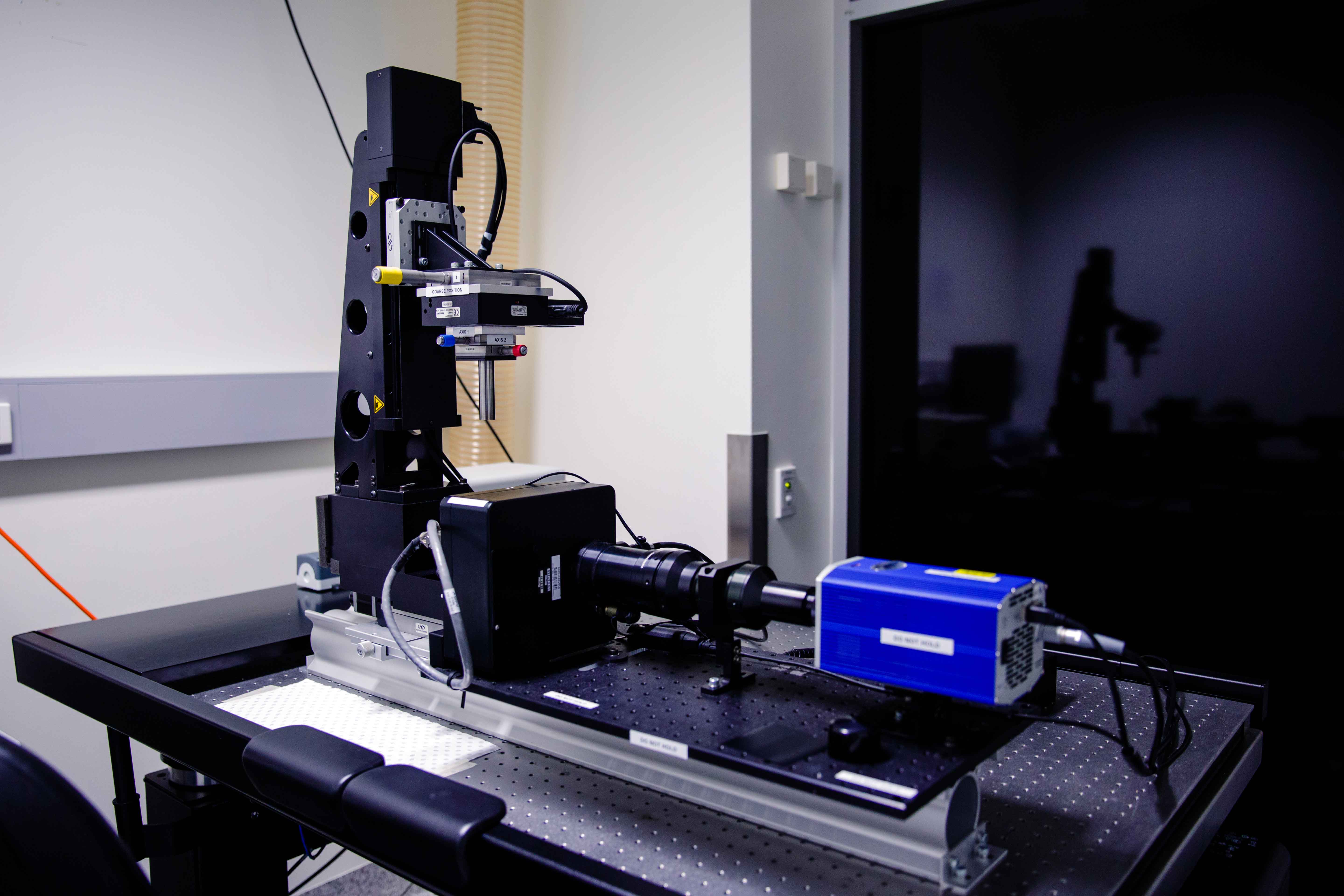 Room 6.033 - Custom Microscope Stand with Photometrics Coolsnap HQ2 CCD camera
Custom widefield microscope with Whitelight Mercury lightsource and CCD camera. Suited for imaging large fixed samples in 3D.
Features
- Fully motorised X-Y-Z stage
Transmitted Light: 100W LED Lamp
Reflected Light: Exfo Xacte 200W Mercury White Lightsource
Objectives
| Position | Objective | Magnification | N.A. | Immersion | W.D. (mm) | Type | Type | Thread |
| 1 | Plan Apochromat | 0.18x | 0.04 | Dry | 50 | |||
| 2 | Plan Apochromat | 0.5x | 0.1 | Dry | 45 | |||
| 3 | Plan Apochromat | 1x | 0.2 | Dry | 40 |
Fluorescent Filter Sets
| Position | Name | Excitation | Dichroic | Emission | Suitable Dyes |
| 1 | DAPI - 5060C | 377/50 | 409LP | 447/60 | DAPI, Hoechst |
| 2 | CFP - 2432C | 438/24 | 458LP | 483/32 | CFP, Cerulean |
| 3 | GFP-3000B | 488/10 | 505LP | 530/60 | GFP, A488 |
| 4 | mCherry LP | 560/20 | 580LP | 585LP | mCherry, Cy3 |
| 5 | - | - | - | ||
| 6 | - | - | - |
Computer
Dell PC - 3GHz Xeon Processor - 8GB RAM - 256GB nVidia Quadro GPU
1TB Raid Array
30" LCD Monitor
Software
OPTiscan
Contacts
Dr James Springfield
Microscopy Facility Manager
+61 7 334 62390
j.springfield@imb.uq.edu.au
Dr Nicholas Condon
CZI Imaging Scientist
+61 7 334 62042
n.condon@imb.uq.edu.au
Dr Mahdie Mollazade
Microscopy Officer
+61 7 334 62042
m.mollazade@uq.edu.au
Mailing address
Advanced Microscopy Facility
Institute for Molecular Bioscience
Level 6N, 306 Carmody Road,
Building 80
University of Queensland
4072, St Lucia,
Queensland, Australia