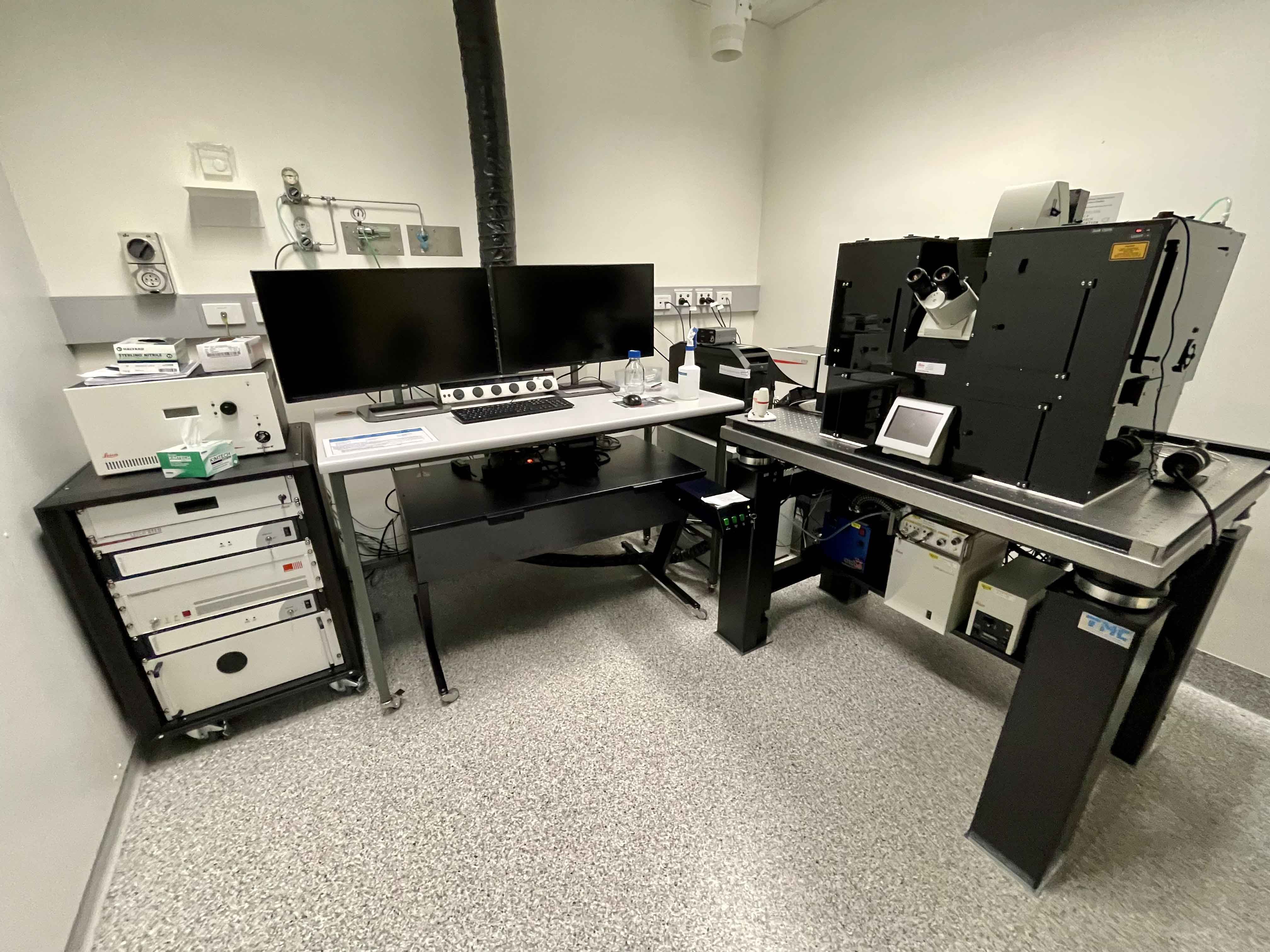
Confocal 1
Inverted Leica STED 3X Confocal with FLIM
.
 Room 6.029a - Leica DMi8 Inverted Microscope Stand with SP8 Galvo and Resonant Confocal Scanners, White Light Laser (WLL), STED 3X super-resolution, and FLIM/FRET FALCON & Lightning Deconvolution
Inverted point-scanning laser confocal microscope with spectral, super resolution and FLIM/FRET detection. Tuneable 470nm - 670nm WLL, 2 x PMT and 2 x HyD detectors. Suited for imaging fixed samples on slides, samples with low fluorescence and live imaging.
Features
- Fully motorised X-Y-Z stage
- High-sensitivity HyD detectors
- CO2 and temperature incubation for live cells and tissue
- Tiled imaging
- Multi-position imaging
- Spectral imaging
- Super-Resolution Imaging with 592nm, 660nm and 775nm STED lasers
- Fast Resonant Imaging
- FLIM/FRET Imaging with WLL and 440nm pulsed lasers
- Adaptive focus control
Transmitted Light: Leica LED Lamp
Reflected Light: Leica EL6000 120W White Light Lamp
Installed in early 2017 is the Leica STED 3X Super Resolution Microscope, which provides multicolour super-resolution imaging of cells. It is the only microscope of its type in Queensland capable of resolving the interactions between single molecules within a living cell in real time. The system comes complete with incubation for live imaging, a resonant scan head capable of 8KHz scan speeds, the super-resolution STED module and also Fluorescent Lifetime Imaging Microscopy (FLIM) capabilities making it a truly versatile and powerful imaging system.
“The STED microscope allows us to look at cells with ultra high magnification in three dimensions. We can reconstruct cancer tissues or small animals, like fish or fly’s, in 3D. It gives us a whole new way to look at cells in health and in disease.” Professor Jenny Stow, Leader The Stow Group - Protein Trafficking and Inflammation.
.
Condensor
| Name | N.A. | W.D. mm | Position 1 | Position 2 | Position 3 | Position 4 | Position 5 | Position 6 |
| Universal LWD | 0.55 | 28 | - | K3 | K6 | K7 | - | - |
Objectives
| Position | Objective | Magnification | N.A. | Immersion | W.D. (mm) | Type | Type | Thread |
| 1 | HC Plan Apochromat | 40x | 1.1 | Water | 0.66 | |||
| 2 | HC Plan Apochromat | 10x | 0.4 | Dry | 2.2 | |||
| 3 | HC Plan Apochromat | 100x | 1.4 | Oil | 0.13 | |||
| 4 | HC Plan Apochromat* | 93x | 1.3 | Glyc | 0.3 | |||
| 5 | - | - | - | - | - | |||
| 6 | HC Plan Apochromat | 40x | 0.95 | Dry | 0.17 |
* Note: The 93x objective also comes with a motorised correction collar.
Fluorescent Filter Sets for Locating through the Eyepiece
| Position | Name | Excitation | Dichroic | Emission | Suitable Dyes |
| 1 |
DAPI |
350/50 |
400 |
460/50 |
DAPI, Hoechst |
| 2 |
GFP |
470/40 |
495 |
525/50 |
EGFP, A488 |
| 3 | Analyzer block | ||||
| 4 | |||||
| 5 |
Rhod LP |
540/45 |
580 |
590 LP |
Texas Red, Cherry |
| 6 | YFP | 500/20 | 515 | 535/30 | YFP |
Lasers
|
Laser |
Wavelength |
Laser Power |
BFP Power* |
Suitable Dyes |
|
405 |
405nm |
50mW |
- |
DAPI, Hoechst |
|
440 pulsed |
440nm |
- |
- |
CFP FLIM/FRET |
|
442 |
442nm | 40mW | - | CFP |
| WLL |
470nm-670nm |
1.5mW |
- |
Visible Dyes |
|
92 STED |
592nm |
1.5W |
- |
Star 488, Oregeon Green |
|
660 STED |
660nm |
1.5W |
- |
Star 470sx, A546 |
|
775 STED |
775nm |
1.5W |
- |
SiR |
*BFP = Back Focal Plane of Objective
Computer
HP Z640 PC - 3GHz Xeon Processor - 64GB RAM - 24GB nVidia Quadro M6000 GPU
3TB Raid Array
2 x 30" LCD Monitor
Software
Leica LASX Software
Co-localisation, 3D visualisation, LAS Navigator, FRAP, FRET, FALCON FLIM and LIGHTNING Deconvolution.
Accessories
Incubation chamber with CO2 and Temperature control
Contacts
Dr James Springfield
Microscopy Facility Manager
+61 7 334 62390
j.springfield@imb.uq.edu.au
Dr Nicholas Condon
CZI Imaging Scientist
+61 7 334 62042
n.condon@imb.uq.edu.au
Dr Mahdie Mollazade
Microscopy Officer
+61 7 334 62042
m.mollazade@uq.edu.au
Mailing address
Advanced Microscopy Facility
Institute for Molecular Bioscience
Level 6N, 306 Carmody Road,
Building 80
University of Queensland
4072, St Lucia,
Queensland, Australia
