Confocal 5
Confocal 6
Zeiss LSM 990 BiG
.
Room 6.019A - Zeiss Axio Observer Inverted Microscope Stand with LSM 990 Confocal Scanner with BiG2 detectors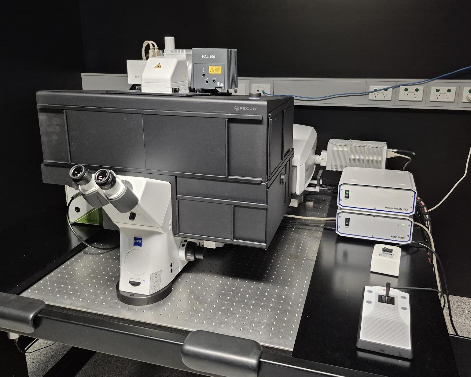
Inverted point-scanning laser confocal microscope. Dedicated BiG2 (GaAsP) detectors for increased sensitivity. Suited for imaging fixed samples on slides, samples with low fluorescence and live imaging.
Features
- Fully motorised X-Y-Z stage
- High-sensitivity BiG detectors (GFP/mCherry and CFP/YFP)
- CO2 and temperature incubation for live cells and tissue
- Tiled imaging
- Multi-position imaging
Transmitted Light: Zeiss 100W Halogen Lamp
Reflected Light: Exfo Xcite Xylis II LED System
.
Condensor
| Name | N.A. | W.D. mm | Position 1 | Position 2 | Position 3 | Position 4 | Position 5 | Position 6 |
| Universal LWD | 0.55 | 26 | - | - | - | - | - | - |
Objectives
| Position | Objective | Magnification | N.A. | Immersion | W.D. (mm) | Type | Type | Thread |
| 1 | Plan Apochromat | 10x | 0.3 | Dry | 2.1 | - | - | M27 |
| 2 | Plan Apochromat | 20x | 0.8 | Dry | 0.55 | - | - | M27 |
| 3 | EC Plan-Neofluar | 40x | 0.75 | Dry | 0.71 | - | - | M27 |
| 4 | LD C-Apochromat | 40x | 1.1 | Water | 0.62 | - | - | M27 |
| 5 | Plan Apochromat | 63x | 1.4 | Oil | 0.19 | - | - | M27 |
| - | - | - | - | - | - | - | - | - |
Fluorescent Filter Sets
| Position | Name | Excitation | Dichroic | Emission | Suitable Dyes |
| 1 | - | - | - | - | - |
| 2 | Zeiss #96 HE | 390/40 | 420 | 450/40 | DAPI |
| 3 | Zeiss #38 HE | 470/40 | 495 | 525/50 | eGFP, FITC |
| 4 | Zeiss #43 HE | 545/25 | 570 | 605/70 | mCherry, Texas Red |
| 5 | - | - | - | - | - |
| 6 | - | - | - | - | - |
Lasers
Wavelength | Laser Type | Minimum Laser Power | Suitable Dyes |
405nm | Diode | 15 mW | DAPI, Hoechst |
445nm | Diode | 10 mW | CFP |
488nm | Diode | 13 mW | GFP, A488 |
514nm | Diode | 13 mW | YFP |
561nm | Diode | 13 mW | mCherry, Cy3 |
594nm | Diode | 4 mW | A594 |
639nm | Diode | 10 mW | Cy5, A647 |
*BFP = Back Focal Plane of Objective
Computer
HP Z8
Software
Zeiss Zen Blue 3.12
Accessories
Incubation chamber with CO2 and Temperature control
Contacts
Dr James Springfield
Microscopy Facility Manager
+61 7 334 62390
j.springfield@imb.uq.edu.au
Dr Nicholas Condon
CZI Imaging Scientist
+61 7 334 62042
n.condon@imb.uq.edu.au
Dr Mahdie Mollazade
Microscopy Officer
+61 7 334 62042
m.mollazade@uq.edu.au
Mailing address
Advanced Microscopy Facility
Institute for Molecular Bioscience
Level 6N, 306 Carmody Road,
Building 80
University of Queensland
4072, St Lucia,
Queensland, Australia