Under the microscope: Unearthing the Soils for Science gallery

What’s lurking outside your house?
Get a glimpse of the weird and wonderful microbes burrowed in backyards across the country on the fascinating Soils for Science image gallery.
Soils for Science, a ground-breaking citizen science initiative from The University of Queensland (UQ)'s Institute for Molecular Bioscience (IMB), has collected thousands of soil samples from everyday Australians to help scientists search for new antibiotics to fight drug-resistant infections, known as superbugs.
 The online image gallery provides a fascinating, up-close insight into what IMB researchers see when they study the samples under the microscope.
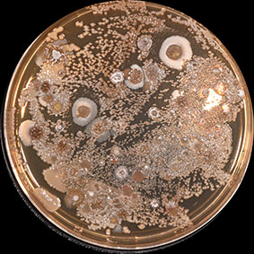
High-resolution images are taken of each sample at various stages of microbial growth, revealing how microbes such as bacteria and fungi interact when they begin to compete with one another.
The gallery images can be explored using the microscope tool to study the samples at up to 32x magnification.
Participants who submit a soil sample can use their unique code to search the gallery for their sample and examine its microbial activity.
The meticulous process of analysing and scanning the samples means it may take a couple of months for images to be uploaded and available to view in the gallery.
Visit the Soils for Science gallery to look at some of IMB’s favourite images and keep checking to see when your microbes are available to view.
IMB Research Fellow Dr Zeinab Khalil is analysing and studying the Soils for Science samples, and reveals how to tell bacteria from fungi, what happens when they interact, and the things scientists look for under the microscope.
Time to get cultured
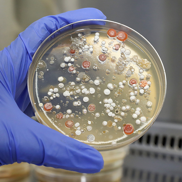
When a Soils for Science sample arrives at IMB’s laboratories, it undergoes a process of analysis to determine what types of microbes, such as bacteria and fungi, are present.
A portion of the soil sample is mixed with sterile water then placed in a petri dish holding a growth medium consisting of agar jelly and other nutrients where cells can be cultured.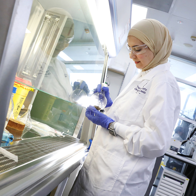
From there, the plate is incubated at 27C for seven to 10 days with continuous supervision, during which cultures begin to form. Following that, IMB scientists such as Dr Khalil examine what is growing on the plate.
“We can see bacteria and fungus growing either close to each other or far away from each other,” Dr Khalil said.
“Once the microbes reveal themselves on the agar plate, it is now the time to start identifying them and this is when we start to check under the microscope.”
Microbial cultures can change dramatically when they grow on an agar plate depending on the media and nutrients added, presenting in various colours and shapes.
“Lots of changes can occur and also be observed when two microbes start to compete with each other,” Dr Khalil said.
“We can also sometimes start to see the production of a pigment that is produced by one microbe, which in turn will inhibit the other microbe from growing.”
By observing the shape of the cultures, researchers can determine whether the microbe they are dealing with is bacteria or fungus.
“Bacteria can be found to be either round (cocci), spiral (spirillum and spirochetes) or rod-shaped (bacillus),” she said.
“While fungi are found to have varying shapes, most are in the form of a thread-like structure called hyphae.”
War of the microscopic worlds
Bacteria and fungus deploy an arsenal of chemical weaponry when fighting over territory, attempting to impede and even destroy one another.
Within these microscopic weapons systems and defence mechanisms are the hidden secrets of nature IMB researchers want to unlock and use to develop new antibiotics.
The ‘zone of inhibition’ is a circular area that forms around an antibiotic strain where competing microbes are unable to grow, indicating its potential for antifungal or antibacterial properties.
“We investigate the microbial interaction and study the change in the morphology of one microbe when it is attacked by another strain and simultaneously, we look for the production of any pigments,” Dr Khalil said.
 “For example, when bacteria are producing red pigment or yellow pigment that inhibits another fungus to grow, this pigment can be antifungal.
“We sometimes see the opposite when a fungus is producing a chemical or a pigment that can inhibit bacteria; this can be an antibacterial agent and affect the morphology and the shape of the other microbe.”
However, not all defence chemicals are coloured – many can be totally colourless but can still be detected by observing the presence of a zone of inhibition around one of the strains.
“We can have bacteria and a fungus growing next to each other but then we start seeing a totally transparent circle formed around the bacteria, which is stopping the other fungus from coming near it,” Dr Khalil said.
“These are the type of things we look for when we check the samples under the microscope and start acquiring images.”
Always a surprise inside
The adaptable nature of microbes, especially when they compete, means researchers can never be sure what they will discover within these microscopic worlds.
Dr Khalil said she can still be surprised at how some microbes grow and change.
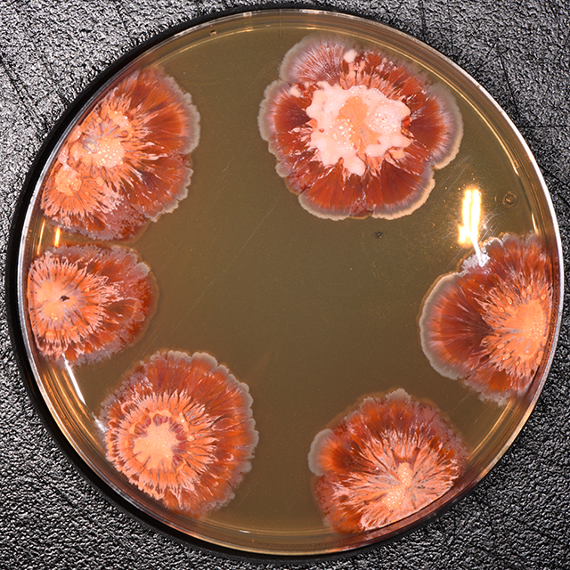 “In one of the soil samples we received, I’ve seen bacteria that grows like a flower on the plate,” she said.
“It looks like a typical jacaranda with different colours of purple growing on the plate and looks really fabulous.”
Studying microorganisms gives researchers profound insight into the inner workings of nature and how it can be harnessed to fight antibiotic-resistant infections.
“It’s interesting to see two microbes fighting with each other, because you can clearly see one is definitely inhibiting the other from growing as if it is trying to protect its territory from the attack by another microbe.”
“It gives us hope that we can find a new medicine in this type of interaction,” Dr Khalil said.
Soils for Science aims to collect 100,000 samples and build a vast library that will sustain antibiotic research well into the future.
Each new sample adds to the growing hope that new antibiotics can be developed to combat drug-resistant superbugs.
“From my perspective, I think every sample is unique,” Dr Khalil said.
“Definitely, each sample will show the growth of some microbes but which one has the potential to produce new antibiotics remains to be seen.
“Soils for Science is a great initiative and a really great opportunity to bring together the public and the scientists, hand-in-hand, to solve a common problem and also to deliver new and improved antibiotics to safeguard future generations.”

Help us find the next antibiotic