Deconvolution Microscopes
Wide-field microscopy involves illumination of the whole sample, and relies on the optical characteristics of the imaging system to capture a single focal plane. The limitation of these kinds of imaging systems is that often, out of focus light (from differing sample planes) are captured in your images, resulting in image-blur. This image blur will lower your signal to noise, which is the contrast of your images. One way to overcome this is by using specialised optics with very narrow depths of field (High NA objectives) coupled with computational algorithms that can correct for out of focus light, this process is called deconvolution.
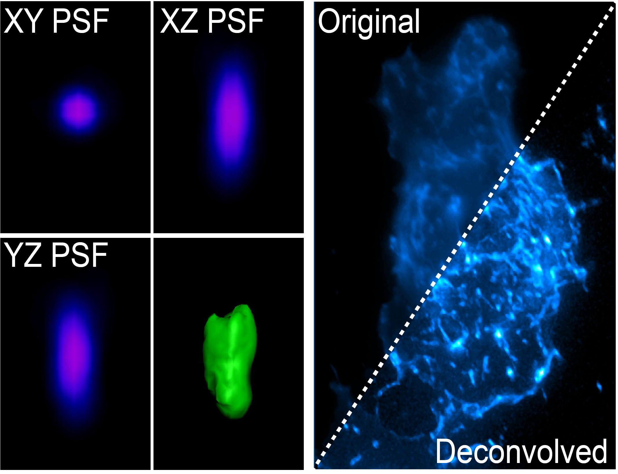
The light coming from your sample scatters (diffraction) in all directions when illuminated, and this can be affected by coverslip thickness, sample mounting medium and the many components of the microscope between your sample and the camera. The way that the light diffracts can be characterised by the point spread function (PSF) whereby a single point of light of known size (fluorescent bead) is captured in xyz and shows the scattering behaviour of light in all dimensions for that particular optical system. The PSF is then used to reverse the effects of out of focus light by correcting for these known distortions in all dimensions.
The PSF can be generated for your particular sample using a fluorescent bead that is smaller than the smallest resolvable structure by the particular imaging system, or it can be theoretically generated using known algorithms. Two main deconvolution algorithms exist, the Richardson-Lucy and Wiener deconvolution algorithms. Deconvolution is an iterative process which is cycled on your images. Deconvolution is a post processing method, and is available either on the microscope (Deltavision, Nikon Deconvolution Microscope) or via software (Analysis Computers, Microvolution, AutoQuant).
Deltavision
Resource Links:
- Deltavision Training Manual
- Deltavision Filter Chart
- Training Videos
- Microscope location - Room 6.025b
- Make a Booking


Room 6.025b - Olympus IX80 Inverted Microscope Stand with Photometrics Coolsnap HQ2 camera
Inverted widefield microscope with Lumencor 7 line LED lightsource, high speed filter wheels and CCD camera. Suited for imaging fixed samples on slides, samples with low fluorescence and live imaging.
Features Include:
- Fully motorised X-Y-Z stage
- High Speed Z-drive for rapid Z-stacks
- CO2 and temperature incubation for live cells and tissue
- Tiled imaging
- Multi-position imaging
Transmitted Light: 100W LED Lamp
Reflected Light: Lumencor Spectra LED Lightsource
Condensor:
Name
N.A.
W.D. mm
Position 1
Position 2
Position 3
Position 4
Position 5
Position 6
Universal LWD
0.45
54
Open
PhL
Ph1
Ph2
DIC I
DIC II
Objectives:
Position
Objective
Magnification
N.A.
Immersion
W.D. (mm)
Type
Type
Thread
1
U-Apochromat
40x
1.35
Oil
-
DIC
M27
2
Plan Apochromat
60x
1.35
Oil
-
DIC
M27
3
UPLS-Apochromat
100x
1.4
Oil
-
DIC
M27
4
-
-
-
-
-
-
5
-
-
-
-
-
-
6
-
-
-
-
-
-
Fluorescent Filter Sets:
Position
Name
Excitation
Dichroic
Emission
Suitable Dyes
1
DAPI
360/40
Quad Band
457/40
DAPI, Hoechst
2
FITC
490/20
Quad Band
526/38
EGFP, Alexa Fluor 488
3
TRITC
555/28
Quad Band
617/63
mCherry, Alexa Fluor 555
4
Cy5
640/20
Quad Band
685/40
Cy5, Alexa Fluor 647
LEDs:
Laser
Wavelength
Laser Power
BFP Power*
Suitable Dyes
390
390nm
20mW
4.5mW
DAPI / Hoechst
440
440nm
5mW
1mW
CFP
485
485nm
20mW
8mW
GFP / A488
514
514nm
3mW
1mW
YFP
563
563nm
10mW
5mW
mCherry / Cy3
594
594nm
5mW
1mW
A594
640
640nm
5mW
2mW
Cy5 / A647
*BFP = Back Focal Plane of Objective
Computer:
Dell PC - 3GHz Xeon Processor - 32GB RAM - 1GB nVidia Quadro GPU
1TB Raid Array
30" LCD Monitor
Software:
Softworx
Accessories:
Incubation chamber with CO2 and Temperature control
Resource Links:
- Deltavision Training Manual
- Deltavision Filter Chart
- Training Videos
- Microscope location - Room 6.025b
- Make a Booking
Room 6.025b - Olympus IX80 Inverted Microscope Stand with Photometrics Coolsnap HQ2 camera
Inverted widefield microscope with Lumencor 7 line LED lightsource, high speed filter wheels and CCD camera. Suited for imaging fixed samples on slides, samples with low fluorescence and live imaging.
Features Include:
- Fully motorised X-Y-Z stage
- High Speed Z-drive for rapid Z-stacks
- CO2 and temperature incubation for live cells and tissue
- Tiled imaging
- Multi-position imaging
Transmitted Light: 100W LED Lamp
Reflected Light: Lumencor Spectra LED Lightsource
Condensor:
Name | N.A. | W.D. mm | Position 1 | Position 2 | Position 3 | Position 4 | Position 5 | Position 6 |
Universal LWD | 0.45 | 54 | Open | PhL | Ph1 | Ph2 | DIC I | DIC II |
Objectives:
Position | Objective | Magnification | N.A. | Immersion | W.D. (mm) | Type | Type | Thread |
1 | U-Apochromat | 40x | 1.35 | Oil | - | DIC | M27 | |
2 | Plan Apochromat | 60x | 1.35 | Oil | - | DIC | M27 | |
3 | UPLS-Apochromat | 100x | 1.4 | Oil | - | DIC | M27 | |
4 | - | - | - | - | - | - | ||
5 | - | - | - | - | - | - | ||
6 | - | - | - | - | - | - |
Fluorescent Filter Sets:
Position | Name | Excitation | Dichroic | Emission | Suitable Dyes |
1 | DAPI | 360/40 | Quad Band | 457/40 | DAPI, Hoechst |
2 | FITC | 490/20 | Quad Band | 526/38 | EGFP, Alexa Fluor 488 |
3 | TRITC | 555/28 | Quad Band | 617/63 | mCherry, Alexa Fluor 555 |
4 | Cy5 | 640/20 | Quad Band | 685/40 | Cy5, Alexa Fluor 647 |
LEDs:
Laser |
Wavelength |
Laser Power |
BFP Power* |
Suitable Dyes |
390 |
390nm |
20mW |
4.5mW |
DAPI / Hoechst |
440 |
440nm |
5mW |
1mW |
CFP |
485 |
485nm |
20mW |
8mW |
GFP / A488 |
514 |
514nm |
3mW |
1mW |
YFP |
563 |
563nm |
10mW |
5mW |
mCherry / Cy3 |
594 |
594nm |
5mW |
1mW |
A594 |
640 |
640nm |
5mW |
2mW |
Cy5 / A647 |
*BFP = Back Focal Plane of Objective
Computer:
Dell PC - 3GHz Xeon Processor - 32GB RAM - 1GB nVidia Quadro GPU
1TB Raid Array
30" LCD Monitor
Software:
Softworx
Accessories:
Incubation chamber with CO2 and Temperature control
Nikon Deconvolution
Resource Links:
- Nikon Filter Chart
- Training Videos
- Microscope location - Room 6.025c
- Make a Booking


Room 6.025c - Nikon Ti-E Inverted Microscope Stand with Hamamatsu Flash 4.0 sCMOS camera
Inverted widefield microscope with Lumencor 7 line LED lightsource, high speed filter wheels and CMOS camera. Suited for imaging fixed samples on slides, samples with low fluorescence and live imaging.
Features Include:
- Fully motorised X-Y-Z stage
- Piezo Z-drive for rapid Z-stacks
- CO2 and temperature incubation for live cells and tissue
- Tiled imaging
- Multi-position imaging
- Transmitted Light: Sutter 100W LED Lamp
Reflected Light: Lumencor Spectra LED Lightsource
Condensor:
Name
N.A.
W.D. mm
Position 1
Position 2
Position 3
Position 4
Position 5
Position 6
Universal LWD
0.45
54
-
-
-
-
-
-
Objectives:
Position
Objective
Magnification
N.A.
Immersion
W.D. (mm)
Type
Type
Thread
1
Plan Apochromat
4x
0.2
Dry
-
-
M27
2
Plan Apochromat
10x
0.45
Dry
4
DIC
M27
3
Plan Apochromat
20x
0.75
Dry
1
DIC
M27
4
Plan Apochromat
40x
0.95
Dry
0.25
DIC
M27
5
S Fluor
40x
1.30
Oil
0.22
DIC
M27
6
Plan Apochromat
60x
1.4
Oil
0.13
DIC
M27
Fluorescent Filters:
Excitation
Dichroic
Emission
390/18
410/504/582/669
440/521/607/700
438/24
462/523
440/40
485/20
505/606
479/40
513/17
436/514/604
525/30
560/25
488LP
542/27
585/29
631/36
650/13
607/36
692/40
488LP
LEDs:
Designation
Wavelength
Power
BFP Power*
Suitable Dyes
390
390nm
-
-
DAPI / Hoechst
440
440nm
-
-
CFP
485
485nm
-
-
GFP / A488
514
514nm
-
-
YFP
563
563nm
-
-
mCherry / Cy3
594
594nm
-
-
A594
640
640nm
-
-
Cy5 / A647
*BFP = Back Focal Plane of Objective
Computer:
Dell PC - 3GHz Xeon Processor - 32GB RAM - 1GB nVidia Quadro GPU
3TB Raid Array
30" LCD Monitor
Software:
NIS Elements 4.3
Accessories:
Incubation chamber with CO2 and Temperature control
Resource Links:
- Nikon Filter Chart
- Training Videos
- Microscope location - Room 6.025c
- Make a Booking
Room 6.025c - Nikon Ti-E Inverted Microscope Stand with Hamamatsu Flash 4.0 sCMOS camera
Inverted widefield microscope with Lumencor 7 line LED lightsource, high speed filter wheels and CMOS camera. Suited for imaging fixed samples on slides, samples with low fluorescence and live imaging.
Features Include:
- Fully motorised X-Y-Z stage
- Piezo Z-drive for rapid Z-stacks
- CO2 and temperature incubation for live cells and tissue
- Tiled imaging
- Multi-position imaging
- Transmitted Light: Sutter 100W LED Lamp
Reflected Light: Lumencor Spectra LED Lightsource
Condensor:
Name | N.A. | W.D. mm | Position 1 | Position 2 | Position 3 | Position 4 | Position 5 | Position 6 |
Universal LWD | 0.45 | 54 | - | - | - | - | - | - |
Objectives:
Position | Objective | Magnification | N.A. | Immersion | W.D. (mm) | Type | Type | Thread |
1 | Plan Apochromat | 4x | 0.2 | Dry | - | - | M27 | |
2 | Plan Apochromat | 10x | 0.45 | Dry | 4 | DIC | M27 | |
3 | Plan Apochromat | 20x | 0.75 | Dry | 1 | DIC | M27 | |
4 | Plan Apochromat | 40x | 0.95 | Dry | 0.25 | DIC | M27 | |
5 | S Fluor | 40x | 1.30 | Oil | 0.22 | DIC | M27 | |
6 | Plan Apochromat | 60x | 1.4 | Oil | 0.13 | DIC | M27 |
Fluorescent Filters:
Excitation | Dichroic | Emission |
390/18 | 410/504/582/669 | 440/521/607/700 |
438/24 | 462/523 | 440/40 |
485/20 | 505/606 | 479/40 |
513/17 | 436/514/604 | 525/30 |
560/25 | 488LP | 542/27 |
585/29 | 631/36 | |
650/13 | 607/36 | |
692/40 | ||
488LP |
LEDs:
Designation |
Wavelength |
Power |
BFP Power* |
Suitable Dyes |
390 |
390nm |
- |
- |
DAPI / Hoechst |
440 |
440nm |
- |
- |
CFP |
485 |
485nm |
- |
- |
GFP / A488 |
514 |
514nm |
- |
- |
YFP |
563 |
563nm |
- |
- |
mCherry / Cy3 |
594 |
594nm |
- |
- |
A594 |
640 |
640nm |
- |
- |
Cy5 / A647 |
*BFP = Back Focal Plane of Objective
Computer:
Dell PC - 3GHz Xeon Processor - 32GB RAM - 1GB nVidia Quadro GPU
3TB Raid Array
30" LCD Monitor
Software:
NIS Elements 4.3
Accessories:
Incubation chamber with CO2 and Temperature control
OBS


Room 6.018 - Olympus IX81 Inverted Microscope Stand with Hamamatsu Orca CCD camera
Inverted widefield microscope with Xenon lightsource, high speed filter wheels and CCD camera. Suited for imaging fixed samples on slides, samples with low fluorescence and live imaging.
Features Include:
- Fully motorised X-Y-Z stage
- CO2 and temperature incubation for live cells and tissue
- Tiled imaging
- Multi-position imaging
Transmitted Light: Olympus 100W Halogen Lamp
Reflected Light: Olympus MT20 Xenon White Lightsource
Condensor:
Name
N.A.
W.D. mm
Position 1
Position 2
Position 3
Position 4
Position 5
Position 6
Universal LWD
0.45
54
Open
PhL
Ph1
Ph2
DIC I
DIC II
Objectives:
Position
Objective
Magnification
N.A.
Immersion
W.D. (mm)
Type
Type
Thread
1
Plan Apochromat
10x
0.4
Dry
-
DIC
M27
2
Plan Apochromat
20x
0.75
Dry
-
DIC
M27
3
Plan Apochromat
40x
1.0
Oil
-
DIC
M27
4
Plan Apochromat
60x
1.35
Oil
-
DIC
M27
Fluorescent Filter Sets:
Position
Name
Excitation
Dichroic
Emission
Suitable Dyes
1
GFP/RFP
S500/20
dual band
dual band
GFP, RFP, mCherry
2
CFP/YFP
S430/25
dual band
dual band
CFP, YFP
3
S565/55
4
S470/30
Computer:
Dell PC - 3GHz Xeon Processor - 8GB RAM - 256GB nVidia Quadro GPU
1TB Raid Array
30" LCD Monitor
Software:
Cell^R
Accessories:
Incubation chamber with CO2 and Temperature control
Room 6.018 - Olympus IX81 Inverted Microscope Stand with Hamamatsu Orca CCD camera
Inverted widefield microscope with Xenon lightsource, high speed filter wheels and CCD camera. Suited for imaging fixed samples on slides, samples with low fluorescence and live imaging.
Features Include:
- Fully motorised X-Y-Z stage
- CO2 and temperature incubation for live cells and tissue
- Tiled imaging
- Multi-position imaging
Transmitted Light: Olympus 100W Halogen Lamp
Reflected Light: Olympus MT20 Xenon White Lightsource
Condensor:
Name | N.A. | W.D. mm | Position 1 | Position 2 | Position 3 | Position 4 | Position 5 | Position 6 |
Universal LWD | 0.45 | 54 | Open | PhL | Ph1 | Ph2 | DIC I | DIC II |
Objectives:
Position | Objective | Magnification | N.A. | Immersion | W.D. (mm) | Type | Type | Thread |
1 | Plan Apochromat | 10x | 0.4 | Dry | - | DIC | M27 | |
2 | Plan Apochromat | 20x | 0.75 | Dry | - | DIC | M27 | |
3 | Plan Apochromat | 40x | 1.0 | Oil | - | DIC | M27 | |
4 | Plan Apochromat | 60x | 1.35 | Oil | - | DIC | M27 |
Fluorescent Filter Sets:
Position | Name | Excitation | Dichroic | Emission | Suitable Dyes |
1 | GFP/RFP | S500/20 | dual band | dual band | GFP, RFP, mCherry |
2 | CFP/YFP | S430/25 |
dual band |
dual band |
CFP, YFP |
3 | S565/55 | ||||
4 | S470/30 |
Computer:
Dell PC - 3GHz Xeon Processor - 8GB RAM - 256GB nVidia Quadro GPU
1TB Raid Array
30" LCD Monitor
Software:
Cell^R
Accessories:
Incubation chamber with CO2 and Temperature control
Lattice Lightsheet Microscope
Room 6.019B
The Lattice Lightsheet microscope is an advanced live cell microscope developed by Nobel Prize winner Eric Betzig in 2014. This microscope is extremely gentle on cells allowing for very long acquisitions, as well as being super-fast, (capturing full 3D volumes in under a second).
Highlights
- Very High spatial resolution (230 nm, 230 nm, 300 nm, xyz)
- Very High Temporal resolution (100 slices / second)
- Very low photo-toxicity
- Opportunity to generate very rare data (very few working systems world wide)
- 4 Individual lasers (440 nm (CFP), 488 nm (GFP), 560 nm (RFP), 640 nm (Far Red))
Challenges
- Massive data sets are generated by this system (terabytes)
- Visualisation and analysis software solutions are not equipped for large volumes of LLSM data, instead MIPS are generated for indexing of files, and finding sections of movies for further analysis. Large collaborations are underway to develop tools to fix this.
- This system is not a ‘turnkey solution’ like our Zeiss systems, therefore the software is not as refined, and often many alignments are required to optimise the imaging output.
Sample Preparation
- This microscope takes 5 mm coverslips (thickness is not important) mounted on the holder shown below.
- The stage can be moved within a 3 x 3 mm region from the centre of the coverslip
- Samples must be adhered to the coverslip (for non-single cell imaging please consult with facility staff)
- Samples are imaged with water dipping lens’ with your sample in a 8 mL water bath (take this into account if your require stimulants/drugs in your imaging media)
- The stage/water-bath is heated, however CO2 is not available while imaging.
- The LLSM room has an incubator set to 37 oC 5% CO2.

To submit a proposal for Lattice Lightsheet Imaging, please use this form.
Room 6.019B
The Lattice Lightsheet microscope is an advanced live cell microscope developed by Nobel Prize winner Eric Betzig in 2014. This microscope is extremely gentle on cells allowing for very long acquisitions, as well as being super-fast, (capturing full 3D volumes in under a second).
Highlights
- Very High spatial resolution (230 nm, 230 nm, 300 nm, xyz)
- Very High Temporal resolution (100 slices / second)
- Very low photo-toxicity
- Opportunity to generate very rare data (very few working systems world wide)
- 4 Individual lasers (440 nm (CFP), 488 nm (GFP), 560 nm (RFP), 640 nm (Far Red))
Challenges
- Massive data sets are generated by this system (terabytes)
- Visualisation and analysis software solutions are not equipped for large volumes of LLSM data, instead MIPS are generated for indexing of files, and finding sections of movies for further analysis. Large collaborations are underway to develop tools to fix this.
- This system is not a ‘turnkey solution’ like our Zeiss systems, therefore the software is not as refined, and often many alignments are required to optimise the imaging output.
Sample Preparation
- This microscope takes 5 mm coverslips (thickness is not important) mounted on the holder shown below.
- The stage can be moved within a 3 x 3 mm region from the centre of the coverslip
- Samples must be adhered to the coverslip (for non-single cell imaging please consult with facility staff)
- Samples are imaged with water dipping lens’ with your sample in a 8 mL water bath (take this into account if your require stimulants/drugs in your imaging media)
- The stage/water-bath is heated, however CO2 is not available while imaging.
- The LLSM room has an incubator set to 37 oC 5% CO2.
To submit a proposal for Lattice Lightsheet Imaging, please use this form.