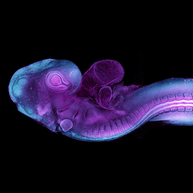
Researchers at IMB have for the first time captured images and video in real time of early embryonic development to understand more about congenital birth defects.
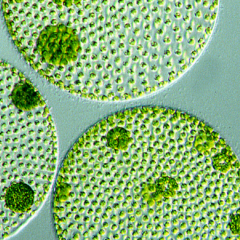
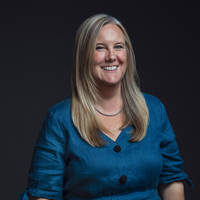
Dr Melanie White and Dr Yanina Alvarez used quail eggs to understand how cells begin to form tissues such as the heart, brain and spinal cord.
3 per cent of babies affected
Dr White said congenital birth defects affect 3 per cent of Australian babies with heart defects the most common and neural tube defects second.
“Because quails grow in an egg, they’re very accessible for imaging and their early development is very similar to a human at the time the embryo implants in the uterus,” Dr White said.
Seeing development in real-time
“For the first time we have seen high-resolution, real-time imaging of important early developmental processes.
“Until now, most of our knowledge of post-implantation development came from studies on static slides, at fixed points in time.”
The IMB researchers have generated quails with a fluorescent protein to reveal the structure, called the actin cytoskeleton, which gives cells shape and facilitates movement.
Cells stick out protusions to move
“When cells migrate during early development, they stick out protrusions called lamellipodia and filopodia like arms that reach out and grab onto surfaces allowing the cells to crawl, or reach other cells to bring them closer together,” Dr White said.
“We were able to image the filopodia from heart stem cells deep inside the embryo as they first made contact by sticking out protrusions and gripping to their surroundings and each other to form the early heart.
“It’s the first time anyone has captured the cell’s actin cytoskeleton facilitating this contact in live imaging.”
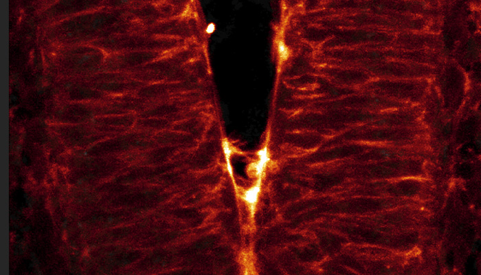
'Zipping' up the neural tube
The researchers also imaged the open edges of the neural tube and it being ‘zipped up’ to begin to form the brain and spinal cord.
“We saw how the cells reached across the open neural tube with their protrusions to contact the opposite side — the more protrusions the cells formed, the faster the tube zipped up,” Dr White said.
“If this process goes awry or is disrupted and the tube doesn’t close properly during the fourth week of human development, the embryo will have brain and spinal cord defects.
Hope for the future
“Our aim is to find proteins or genes that can be targeted in the future or used for screening for congenital birth defects.
“We are very excited at the possibilities that this new quail model now offers to study development in real time.”
The research is published in the Journal of Cell Biology by a team that included IMB's Marise van der Spuy and Jian Xiong Wang.
Rare diseases take centre stage on Rare Disease Day
Dr Melanie White and Dr Anna Lagendijk