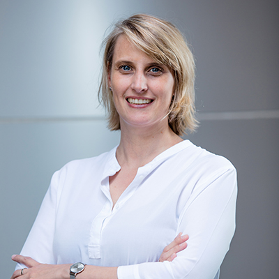
Researcher Profile: Dr Anne Lagendijk
What is the question you are trying to answer?

How is our blood vessel network established during development and how is its function maintained throughout life?
How does your research relate to stroke?
When the cells that make up our blood vessels fail to maintain their shape and function, this can lead to vascular malformations, like Cerebral Cavernous Malformations (CCMs). CCMs arise as clusters of vessels with an enlarged diameter, most often in the brain.
CCM vasculature consists of severely enlarged, thin cells, making these vessels highly compliant and prone to rupture. As a result of rupture there is an increased risk of hemorrhagic stroke, focal neurological defects and seizures.
My lab studies what causes CCM formation at a cellular level in live zebrafish embryos. This live approach is unique and we aim to understand more about the drivers of CCM and more broadly of cell shape maintenance in order to prevent bleedings.
Why is it important to research this area?
This work will gain a deeper understanding of CCM pathogenesis. Furthermore, it will expand our knowledge on key cellular components that more generally underpin vessel integrity and the biology of stroke.
What are the big research questions in stroke?
Which vessels of the brain are prone to rupture?
Is there a role for the vessel environment in susceptibility to stroke?
How do we repair ruptured vessels?
What motivated you to become a researcher?
I was fortunate to get the opportunity to work at the Hubrecht Institute (Utrecht, the Netherlands), a world-renowned institute for Developmental Biology and Stem Cell Research, while finishing my MSc degree. I became inspired by the amazing discoveries around me and wanted to take part in discovery-based science with the aim to uncover more and more of the unknown that will ultimately help us to better understand disease.
How did you get into this area of research?
During my PhD work I stayed at the Hubrecht in the lab of Prof. Jeroen Bakkers and uncovered the function of known and novel genes that are required to make cardiac valves. During my PhD there was an ongoing shift in the field. Where initially we investigated development at a whole tissue level, advances in microscopy allowed us to examine development in greater detail, at a cellular level in live animals. For my postdoctoral work at the IMB I challenged the level of detail even further by making a zebrafish line that allowed us to measure mechanical changes between cells of our vasculature. Cellular forces are known to be essential for cellular adaptation and communication, but investigating the role of forces in a 3D live vasculature had not been done previously. I believe that in order to know how a healthy vessel is maintained and thus prevent stroke, we need to learn how the individual cells within these vessels work and communicate with neighbouring cells and their environment.
Who/What inspires you?
The fact that nothing is unimaginable. During my scientific career there have been two major advances in the field 1) allowing us to modify genomes (CRISPR) and 2) being able to analyse the genetic activity of individual cells (single cell sequencing). Just these two techniques expanded the capacity of our research to a scale that I would not have predicted when I started. We are in a very exciting era in science where we can uncover what keeps individual cells in check which will contribute to the development of more targeted therapies in the future.
What is one thing or fact that amazes you about the body?
It still amazes me that a single fertilised cell can give rise to an immense array of different cell types that together form the various organs of our body, including a fully functional cardiovascular network.
